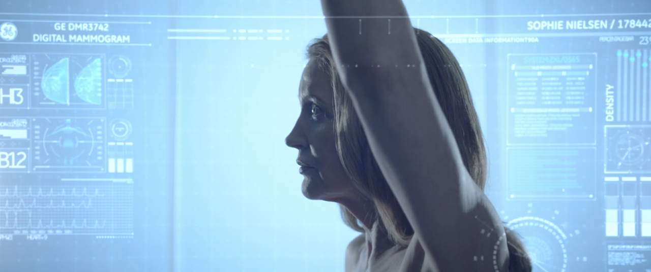
Developments in mammography imaging have changed the way radiologists and oncologists work as well as the lives of the women who have experienced this essential technology. For the first time in 20 years, the U.S. Food and Drug Administration (FDA) has announced new steps it is taking to modernize breast cancer screening in ways that empower the patient and support decision making.1 Changes include modernizing quality standards, and adding new assessment categories and tissue density reporting requirements.1
Advancements in mammography imaging technology are reportedly already positively impacting the health of women of all ages in real ways that improve care delivery and outcomes; they include an increased and improved ability to detect breast cancer at the earliest possible moments in its development, confirmation of the benefits of 3D imaging for early breast cancer detection, and the ability of artificial intelligence (AI) to accurately and reliably classify breast tissue density without the inherent human variabilities.
Digital mammography paved the way for better cancer detection
Advancements in mammogram technology using digital 2D imaging became an accepted standard method for breast cancer screening and detection 15 years ago because of several improvements over screen film mammography, based on a study conducted in the U.K.2 For example, 2D mammography produces higher quality visualizations for detecting calcifications and seeing through denser breast tissue, which often masks the presence of cancer.2 Additionally, digital images can be adjusted on-screen, allowing radiologists to get a better visual.2
The U.K. retrospective study published in Radiology has confirmed the superiority and increased impact of 2D imaging over screen film mammography for cancer detection after analyzing the data of 11.3 million screening exams from 80 facilities--breast cancer is more likely to be identified by digital mammography.2
For women ages 45 to 70, this technological advancement has translated into a 14 percent higher rate of detection for invasive early-stage grade one and grade two breast cancers, according to that same study.2 That increases to a 19 percent higher detection rate during first screening exams for women between the ages of 45 and 52.2 Detecting these cancers early is key to treatment success and survival because they are the disease types most likely to develop into life-threatening illness, particularly when they are not identified, diagnosed, and treated at the earliest possible opportunity.2
Researchers of the retrospective study give full credit for better detection rates to the improvements in technology given no other changes, such as computer-aided detection, were made to the screening program.2 Another significant benefit of 2D mammography for patients is, despite the increase in detection rate, there was no increase in the recall rate for false positives, meaning patients wouldn't undergo unnecessary further screening because of abnormal results.2
3D mammography achieves early detection in older women
According to national statistics half of the more than 250,000 new invasive breast cancer diagnoses estimated annually occur in women age 60 or older with early detection during screening exams providing the best chance for survival.3 Despite this, the benefits of breast cancer screening in women age 65 and older are not embraced by all health professionals.4 For example, the U.S. Preventive Services Task Force recommends a cut-off age of 74.4 In contrast, no age-based recommendations are given by other medical experts.4 Instead, they suggest a patient's personal preferences, state of health, and life expectancy are more appropriate guidelines for making determinations about when to stop screening exams.4
Results from a new retrospective study agree. Mammograms from more than 35,000 women with an average age of 72 who had 2D mammography or 2D plus 3D mammography were analyzed. Although no specific age for stopping breast cancer screening was supported by analysis of the study data,4 the performance metrics for screening are favorable in women 65 years and older and are further improved with digital breast tomosynthesis, or 3D mammography.4
Several advantages of the 3D method were confirmed, such as a reduction in false positives and abnormal interpretations that lead to unnecessary additional screening and worry.4 Tomosynthesis also provided greater value for predicting the likelihood that patients who received a positive screening result would be diagnosed with breast cancer.4 Tomosynthesis also resulted in higher specificity than exams conducted with 2D mammography, meaning there were fewer false positives.4 Additionally, researchers concluded that fewer lymph node-positive cancers were detected in tomosynthesis exams because the presence of cancer was being found at an earlier stage, the ultimate goal of mammography.4
Algorithm categorizes dense tissue in mammograms
Women with dense breast tissue are at such a significantly greater risk for developing cancer that the FDA, along with many individual states, has implemented legislation requiring reporting and notification to patients when their mammograms result in a dense tissue classification.5 This is because tissue density is not only an independent risk factor, but also makes cancer detection more challenging, potentially delaying diagnosis and treatment, all of which impact the ability to achieve positive outcomes and survival.5
A major factor impacting the classification of dense breast tissue is human variability, given the evaluation process relies on radiologists.5 All mammograms must receive a Breast Imaging Reporting and Data System (BI-RADS) breast density rating based on four categories: fatty, scattered fibroglandular, heterogeneously dense, and dense.5 These rating take time to complete, and inherent differences between radiologists, such as experience level, fatigue, and so on, can influence the results.5
Given these factors, physicians from Massachusetts General Hospital (MGH) and researchers from the Massachusetts Institute of Technology focused their efforts using artificial intelligence to build an algorithm that can quickly and accurately assess breast density. They built a deep-learning convolutional neural network (CNN) that was trained and tested on more than 58,000 digital mammograms to differentiate between all four BI-RADS categories as well as experienced radiologists can.7
Once the algorithm was trained and tested, they showed how it could be implemented into clincial practice effectively. They had the algorithm assess nearly 11,000 consecutive mammograms and assign each a density rating before sending it to one of eight radiologists who weren't involved in the training portion of the study. Once the radiologist received the image they could see the algorithm-assigned rating and either accept or reject it.5
The algorithm matched the eight radiologists's decisions 94 percent of the time when it was tested on whether breast tissue was either fatty and scattered, or heterogeneous and dense, and matched the radiologists 90 percent of the time across all four BI-RADS categories.5
As of January 2018, the algorithm has been implemented into MGH's clinical process, and every digital mammogram is automatically assigned a density rating before a radiologist's evaluation, which marks the first time a deep-learning model has been successfully incorporated into a real-time, routine clinical setting.6 And the results suggest the algorithm's ability to predict density category could lead to the standardization and automation of routine breast tissue density assessment.5
Building deep-learning algorithms like this one requires large, mature datasets, which makes it well matched to be used with breast cancer screening because of the vast, advanced, and structured reporting that links breast images like mammograms with outcomes; and this first application is a pivotal technology for the continued development of personalized breast cancer risk assessment.5
REFERENCES:
- FDA Proposes New Rules for Mammography Reporting and Quality Improvement. Imaging Technology News https://www.itnonline.com/content/fda-proposes-new-rules-mammography-reporting-and-quality-improvement Accessed 4/23/2019
- Digital mammography increases breast cancer detection. Medical Express https://medicalxpress.com/news/2018-12-digital-mammography-breast-cancer.html AND study link https://pubs.rsna.org/doi/full/10.1148/radiol.2018181426 Accessed 4/23/2019
- Breast Cancer Facts & Figures 2017-2018. American Cancer Society https://www.cancer.org/content/dam/cancer-org/research/cancer-facts-and-statistics/breast-cancer-facts-and-figures/breast-cancer-facts-and-figures-2017-2018.pdf Accessed 4/23/2019
- Older women benefit significantly when screened with 3-D mammography. Medical Express https://medicalxpress.com/news/2019-04-older-women-benefit-significantly-screened.html AND study link https://pubs.rsna.org/doi/10.1148/radiol.2019181637 Accessed 4/23/2019
- Deep-learning algorithm identifies dense tissue in mammograms. Physics World https://physicsworld.com/a/deep-learning-algorithm-identifies-dense-tissue-in-mammograms/ AND study link https://pubs.rsna.org/doi/10.1148/radiol.2018180694 Accessed 4/23/2019
- Artificial Intelligence Used in Clinical Practice to Measure Breast Density. EurekAlert. https://www.eurekalert.org/pub_releases/2018-10/rson-aiu101018.php Accessed 4/25/2019.
- Mammographic Breast Density Assessment Using Deep Learning: Clinical Implementation. Radiology. https://pubs.rsna.org/doi/10.1148/radiol.2018180694. Accessed May 8, 2019.


