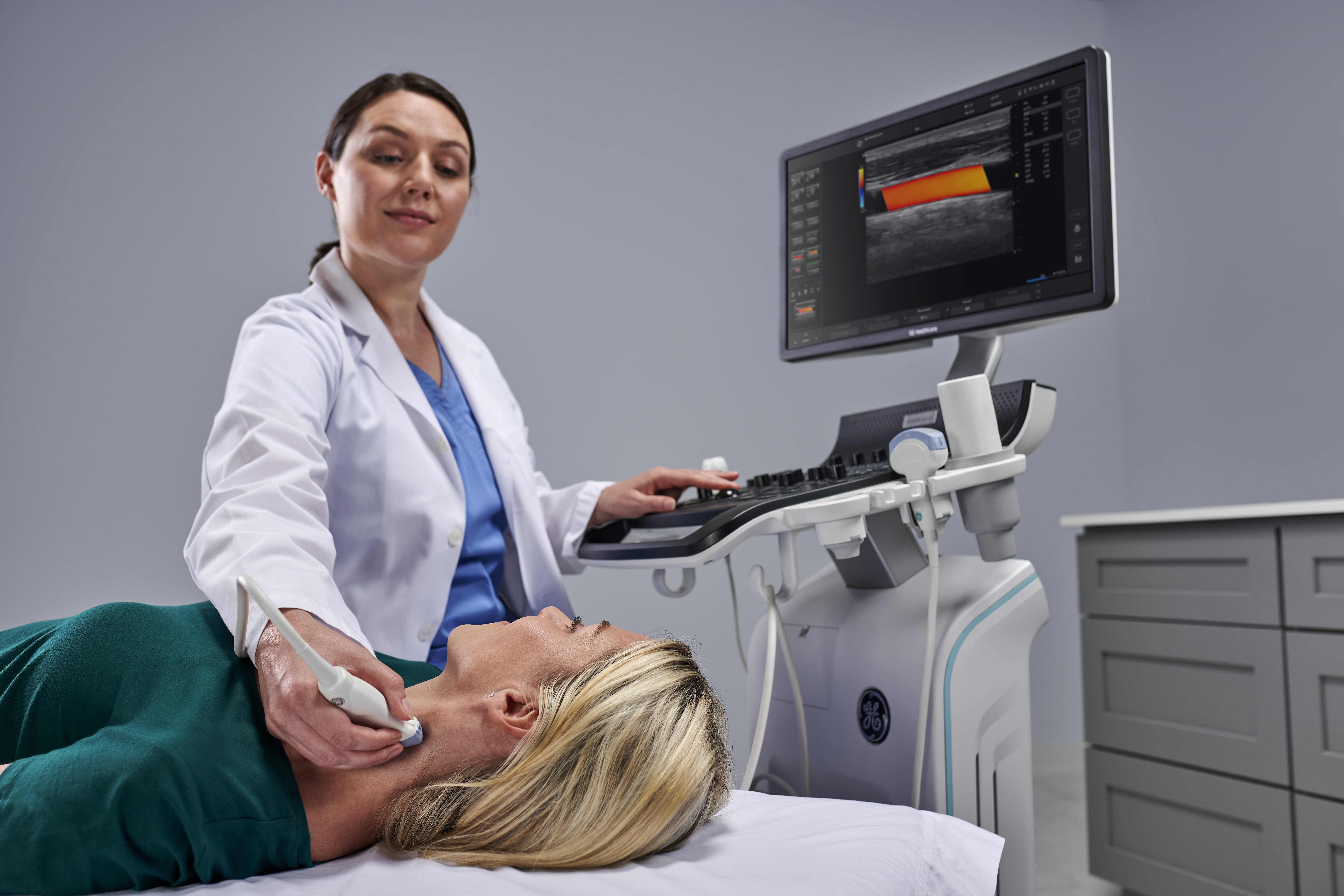
Today's primary care practices are witnessing the dawning of the "age of the ultrasound system." More and more general practitioners (GPs) are performing scans to aid in the diagnosis of a wide range of conditions, from abdominal issues to musculoskeletal (MSK) injuries. A recent targeted research study indicates that nearly half of all GPs are integrating ultrasound into their practices,1 supporting its viability as a diagnostic solution that improves treatment speed and quality and reduces healthcare costs.2
Driving this continued proliferation among GPs is ultrasound's versatility of application and the degree to which it can expand practices' scope of care. The ability to proactively diagnose conditions across so many areas of specialization can 1) help augment care for current patients, 2) position a practice to serve new populations as demographics change, and 3) support expansion to other facilities.
Current ultrasound technology allows GPs to treat patients more comprehensively, in-office, across all stages of life. From early fetal development and dating ultrasounds through adult and geriatric abdominal, cardiac and MSK scans later in life, today's GPs can leverage ultrasound to offer continuous care and diagnostic precision.
Let's examine primary care ultrasound's most significant impacts on health outcomes to date and discuss what this technology's introduction could mean for your practice.
Abdominal ultrasound in primary care
One of the most common uses of ultrasound among GPs is the evaluation of abdominal symptoms.3 In one major study, abdominal imaging accounted for nearly 70% of all scans captured by doctors. GPs can perform abdominal scans to detect and diagnose a number of health issues that originate with abdominal symptoms.
Clinicians in primary care environments also use abdominal scans to detect issues in the liver and gall bladder,4 including fatty liver, hemangiomas, biliary dilatation, choledocholithiasis, cholelithiasis, and renal abnormalities. This improves the odds of early detection, which in turn allows for more proactive care, better patient outcomes, and fewer referrals to external imaging specialists.
Early detection and management of thyroid issues
Ultrasound is also commonly utilized to evaluate thyroid nodules, thyroid size, and thyroid gland abnormalities, supporting diagnoses of disorders such as thyroiditis, goiters, and potential thyroid malignancies. An established and growing body of international and domestic research indicates that the American College of Radiology Thyroid Reporting and Data Systems (ACR®-TIRADS®) classification can assist in thyroid nodule diagnosis and reduce the frequency of unnecessary invasive biopsies.5
While thyroid ultrasound has traditionally been the purview of imaging specialists and endocrinologists, today's more intuitive systems, which offer thyroid-specific productivity tools and other user-friendly features, make scans more manageable for GPs.
The emergence of cardiac ultrasound in primary care
Research also suggests that focused ultrasound can viably detect cardiac abnormalities and improve treatment plan timelines. Cardiac scans provided findings that helped change the predetermined course of care; one study revealed that cardiac ultrasound helped GPs detect aortic stenosis and cardiac failure when it was missed in previous clinical examinations.6
Newer users can also deploy cardiac ultrasound to check ventricular function7 and detect abnormalities (e.g., ventricular hypertension (LVH), valvular insufficiency, hypertrophic cardiomyopathy (HCM), dilated cardiomyopathy (DCM), septal defects, and ejection fraction issues, while also diagnosing heart failure more quickly.8
Breast ultrasound in primary care
As women continue to battle escalating rates of breast cancer,9 early detection is critical to optimizing treatment outcomes, prognosis, and quality of life. Unfortunately, there continue to be gaps in detection for this incredibly time-sensitive condition.10
Just as with thyroid disease, today's most advanced ultrasound systems equip GPs to perform breast scans more confidently, via specialized probes and productivity tools that leverage Breast Imaging Reporting and Data System (BI-RADS®)* criteria.
Maternal-child health scans, now in GPs' hands
Obstetrics represents another application for primary care ultrasound functions. Recent innovations in ultrasound technology enable even newer users to detect early pregnancy, fetal anatomy, growth and measurements, and pelvic and ovarian abnormalities. Automated tools available on some of the latest systems help new ultrasound users simplify standard fetal measurements while optimizing and analyzing color flow and image quality.
Pelvic ultrasound, increasingly common in primary care environments, has become a standard evaluation to assess the uterus, ovaries, fallopian tube, and bladder.10 The right ultrasound system offers features like auto contour measuring or 3D/4D imaging capabilities, which simplify pelvic scans for new users.
Musculoskeletal ultrasound in primary care venues
Musculoskeletal (MSK) scans also reflect the evolution of ultrasound in primary care. This application helps GPs improve the quality of care for conditions like muscle tears, tendonitis, bursitis, joint problems, rheumatoid arthritis, and masses (e.g., tumors or cysts). MSK scans also help guide a wide array of injectable treatments, local anesthetics, corticosteroids, platelet-rich plasma, and more options to treat tendonitis, bursitis, and ganglion cysts.
Primary care ultrasound in vascular medicine
Ultrasound-assisted vascular injections have significantly "democratized" vascular treatment for primary care patients, helping identify peripheral artery disease, assess plaque in the carotid artery, and track blood flow, blockages, and clots that can signal the need for more urgent intervention. The latest ultrasound systems offer the most advanced image optimization tools, helping GPs capture the clearest scan possible while accessing obscure views once considered problematic for newer and even more experienced users.
So, what's behind this evolution?
Advancements in ultrasound technology have unlocked diagnostic possibilities once considered out of the reach of the primary care environment. Scanning functions once relegated to specialists and imaging centers are now available in the comfort and privacy of patients' own doctor's offices.
In fact, the speed of ultrasound expansion may suggest revolution rather than evolution. Not only do scan-assistant tools, automated exam presets, hands-free voice comments, and condition-specific probes that integrate with a single system simplify scans, but more comprehensive education and support tools make it easier and faster for newer users to learn and hone their expertise. Finally, artificial intelligence and automation tools are greatly improving clinical workflow. So, if your primary care practice has been considering offering ultrasound, there's truly no time like the present to select the right system for your patients.
Unlock the full potential of your practice—discover the benefits of integrating primary care ultrasound today.
*BI-RADS and ACR are trademarks of American College of Radiology
Resources:
-
Touhami D, Merlo C, Hohmann J, et al. The use of ultrasound in primary care: longitudinal billing and cross-sectional survey study in Switzerland. BMC Family Practice. 2020;21(1). https://doi.org/10.1186/s12875-020-01209-7
-
Andersen CA, Holden S, Vela J, et al. Point-of-care ultrasound in general practice: a systematic review. Annals of Family Medicine. 2019;17(1), 61–69. https://doi.org/10.1370/afm.2330
-
Alamri AF, Khan I, Baig MI, et al. Trends in ultrasound examination in family practice. Journal of Family & Community Medicine. 2014;21(2),107–111. https://doi.org/10.4103/2230-8229.134767
-
Tollefson B, Hoda N, Fromag G, et al. Bedside gallbladder ultrasound for the primary care physician. J Miss State Med Assoc. 2015 Mar;56(3):64-6. https://pubmed.ncbi.nlm.nih.gov/26050444/
-
Leni D, Seminati D, Fior D, et al. Diagnostic performances of the ACR-TIRADS System in thyroid nodules triage: a prospective single center study. Cancers (Basel). 2021;13(9):2230. doi: 10.3390/cancers13092230.
-
Yates J, Royse CF, Royse C, et al. Focused cardiac ultrasound is feasible in the general practice setting and alters diagnosis and management of cardiac disease. Echo Research and Practice. 2016;3(3), 63–69. https://doi.org/10.1530/ERP-16-0026
-
Mjølstad OC, Snare SR, Folkvord L, et al. (2012). Assessment of left ventricular function by GPs using pocket-sized ultrasound. Family Practice. 2012;29(5), 534–540. https://doi.org/10.1093/fampra/cms009
-
Magelssen MI, Hjorth‐Hansen AK, Andersen GN, et al. (2023). Clinical influence of handheld ultrasound, supported by automatic quantification and telemedicine, in suspected heart failure. Ultrasound in Medicine and Biology. 2023;49(5), 1137–1144. https://doi.org/10.1016/j.ultrasmedbio.2022.12.015
-
Xu S, Murtagh S, Han Y, et al. (2024). Breast cancer incidence among US women aged 20 to 49 years by race, stage, and hormone receptor status. JAMA Network Open. 2024;7(1), e2353331. https://doi.org/10.1001/jamanetworkopen.2023.53331
-
Seely JM. (2023). Progress and remaining gaps in the early detection and treatment of breast cancer. Current Oncology. 2023;30(3), 3201–3205. https://doi.org/10.3390/curroncol30030242
JB31600XX

