The potential offered by emerging medical technologies in cancer care is undeniable. Telehealth, liquid biopsy, next-generation targeted therapies, robotic surgery, radiotheranostics, immuno-oncology are examples of technologies that are reshaping the ways clinicians diagnose and treat cancer. In the reality where it usually takes a long time to change guidelines and implement use in practices, the challenge is finding the best ways to incorporate these new technologies so that patients and oncology programs may benefit sooner. Yet, in fact, according to a recent study published in JAMA Medical News, this process can take as long as 17 years.[i]
Are cancer care programs keeping pace with the explosive growth of data to handle novel technologies? This is one of the key questions clinicians worldwide are trying to find answers to, in addition to how to improve cancer outcomes.
Integrating new technologies into everyday patient care
Integrating technology into care is a complex process that requires time, expertise, and a steadfast commitment to always having the patient’s welfare in focus.
What should clinicians consider when implementing a new technology at their hospital or clinic? Dr. Narayana Subramaniam, Director of Head and Neck Surgery and Oncology, and Director of Clinical Innovation at Sparsh Hospitals in Bengaluru, India, specializes in adapting new technologies for clinical practice. “These technologies are making significant strides in medicine now because the rapid advancement of tech has brought them closer to the doctors, to the patients, making them much more accessible,” he says. “Before, there was a significant entry barrier because of cost, but that’s not an issue anymore.”
He recommends this strategic roadmap: start with a clear clinical problem in mind, evaluate how technology can enhance existing workflows, understand the context of the technological solution, ensure that technology is not a key influencer in a doctor-patient relationship, and prioritize testing and validation to uphold patient trust.

Dr. Narayana Subramaniam is Director of Head and Neck Surgery and Oncology and Director of Clinical Innovation, Sparsh Hospitals, Bengaluru, India. He specializes in adapting new technologies to clinical practice.
How radiomics is impacting cancer careIn addition to thinking through the strategic roadmap on how to integrate these technologies, providers need to also consider artificial intelligence and machine learning, which represent enormous potential in oncology. Radiomics, is a concept which implements machine learning to identify specific features in various imaging modalities data and embedded analytics for diagnostic, prognostic, and treatment planning purposes. It can be used to answer very specific questions and as a triage tool, aid in decision-making by trained radiologists. Radiomics can incorporate a significant amount of data that we, as human beings, don’t perceive, or data that is not available to us, providing an additional layer of data might be very valuable for addressing cancer. This emerging technology is becoming a new chief consideration for providers globally as they consider making significant strides in impacting cancer care.
Deep learning and radiomics algorithms are helping improve accuracy, sensitivity, specific and precision
Another approach that is emerging thanks to researchers at the Karl Landsteiner University of Health Sciences, is the use of machine deep learning, and a convolutional neural network (CNN) for implementing radiomics principles and analyzing imaging data to diagnose specific types of brain tumors.
Most common neoplasms in the brain are represented by gliomas of brain metastases and brain metastases. Precise characterization of the tumor type is pivotal for the success of the treatment. Yet, there may be limitations to what human eye can see in order to differentiate out of conventional MRI data.
In the recent research study from Karl Landsteiner University[ii], research team leader Prof. Andreas Stadlbauer said: “In all important differentiation criteria such as accuracy, sensitivity, specificity and precision, our CNN managed to clearly differentiate the two tumor types.”
AI/ML-aided pathology to gain valuable insights and make more accurate predictions
Another emerging technology using convolutional neural networks for semi-automated cancer diagnoses is AI / DL aided pathology. It’s commonly used today to diagnose cervical, lung, prostate, and oral or head and neck cancer. Morphological features of cancer can be identified from the cells on a slide, in spite of large amounts of variability. In recent years, the deep neural network has shown the same accuracy as clinicians in the diagnosis of cutaneous malignant melanoma[iii] . In the field of ophthalmology, the depth neural network assessment of retinal fundus images shows high sensitivity and specificity, which is conducive to the detection of diabetic retinopathy[iv]. The development trend of DL is gradually applied to new fields, including lung cancer pathology.
With machine learning, we have the potential of revolutionizing the field of healthcare, and particularly pathology, by enabling the analysis of large and complex datasets.
Using different data types, preprocessing techniques, and machine learning algorithms, it is possible to gain valuable insights and make more accurate predictions. With the use of supervised and unsupervised learning methods, a wide range of tasks can now be performed, including classification, clustering, and regression.[v]
Creating patient-specific implants with 3D printing
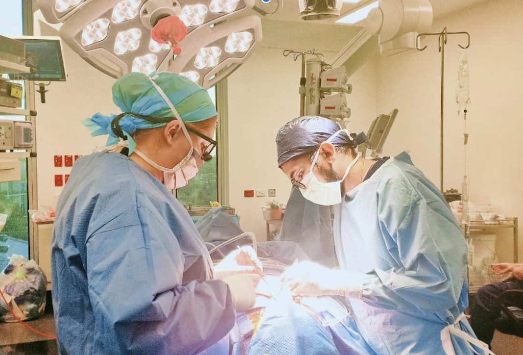
Another example of technology that offers significant potential in cancer care is 3D printing. There’s been a dramatic fall in the cost of medical 3D printing devices, with high-quality printers now accessible at an affordable price point. This means there is no longer a need to mill items like surgical planning guides or implants.
The ability to perform virtual surgical planning and medical three-dimensional (3D) printing has resulted in helping reduce operating times, improve accuracy and efficiency, increase predictability and repeatability, enhance resection and reconstruction accuracy, and facilitate dental rehabilitation and functional jaw reconstructions in head and neck cancers.[vi] With the advancement of these innovations, along with computer-assisted surgery (CAS), head and neck surgery and craniofacial surgery, has been revolutionized.”[vii] Today, surgical uses for 3D printing includes surgical tools and planning guides, made of resin or PMMA (polymethyl methacrylate). Implantable materials can be printed using PEEK (polyetheretherketone), titanium or silicone. Non-surgical applications encompass radiotherapy delivery, bio-scaffolds (using bio-absorbable materials that are replaced over time by the body’s tissues), and drug delivery.
Genetic and molecular testing that leads towards more targeted and personalized treatments
While genetic testing has been around since the mid-1990s, testing for BRCA1 or BRCA2 genes mutations was introduced to identify patients at risk for hereditary breast and ovarian cancer syndrome,[viii] that has led to applications expanding exponentially. Today, it is estimated that approximately 5% to 10% of cancers can be linked to germline pathogenic variants.[viii] There are multiple studies currently underway to prove the value of intensified screening for prostate cancer in men with germline BRCA2, BRCA1, MSH2, and MSH6 mutations, for example, which will lead to more personalized treatment approaches. [viii]
Molecular testing, a cornerstone in modern oncology, opens doors for greater cancer therapy personalization. With an expanding scope, especially in identifying genetic predispositions to certain cancers, molecular testing is now pivotal in tailoring treatment strategies, for example in non-small cell lung cancer.
A recent compelling study by researchers at the University of Liverpool has further expanded the scope of molecular testing. The study focused on identifying lung cancer risk predictively through the analysis of plasma protein biomarkers. Utilizing four distinct machine learning algorithms, the research team successfully pinpointed individuals at heightened risk of developing lung cancer. This was achieved by monitoring the participants over a period, underscoring the prognostic power of these biomarkers. The significance of this study lies in its demonstration of using minimally invasive biomarker testing to not only predict lung cancer but also to potentially prompt earlier detection through personalized risk assessment. [ix]
Technology as a force multiplier
In conclusion, Dr. Subramaniam says, “we need to look at technology as a potential force multiplier that enhances, rather than dramatically alters, established healthcare processes.
While the road to full implementation might be long, the ultimate rewards – a revolution in patient care – make the journey decidedly worthwhile.“ Whether focusing on AI, Machine or Deep Learning, Biomarkers, 3D Printing or other emerging technologies, staying current on the latest innovations and learning how to integrate into practice and demonstrate outcomes that can impact cancer care is at the forefront of how to advance your clinical acumen.
For more information on GE HealthCare Oncology solutions, click here.
References
[i] Rubin R. It Takes an Average of 17 Years for Evidence to Change Practice—the Burgeoning Field of Implementation Science Seeks to Speed Things Up. JAMA. 2023;329(16):1333–1336. doi:10.1001/jama.2023.4387
[ii] Differentiation of Glioblastoma and Brain Metastases by MRI-Based Oxygen Metabolomic Radiomics and Deep Learning. A. Stadlbauer, G. Heinz, F. Marhold, A. Meyer-Bäse, O. Ganslandt, M. Buchfelder & S. Oberndorfer. Metabolites 2022, 12 (12), 1264. https://doi.org/10.3390/metabo12121264
[iii] Esteva A, Kuprel B, Novoa RA, Ko J, Swetter SM, Blau HM. et al. Corrigendum: Dermatologist-level classification of skin cancer with deep neural networks. Nature. 2017;546(7660):686. [PubMed] [Google Scholar]
[iv] Gulshan V, Peng L, Coram M, Stumpe MC, Wu D, Narayanaswamy A. et al. Development and Validation of a Deep Learning Algorithm for Detection of Diabetic Retinopathy in Retinal Fundus Photographs. JAMA. 2016;316(22):2402–10. [PubMed] [Google Scholar]
[v] Artificial intelligence and machine learning overview in pathology & laboratory medicine: A general review of data preprocessing and basic supervised concepts Volume 40, Issue 2, March 2023, Pages 71-87 https://www.sciencedirect.com/science/article/pii/S0740257023000138
[vi] Yang WF, Choi WS, Leung YY, Curtin JP, Du RX, Zhang CY, et al. Three-dimensional printing of patient-specific surgical plates in head and neck reconstruction: A prospective pilot study. Oral Oncol (2018) 78(3):31–6. doi: 10.1016/j.oraloncology.2018.01.005 https://www.frontiersin.org/journals/oncology/articles/10.3389/fonc.2022.960545/full
[vii] Yang WF, Choi WS, Leung YY, Curtin JP, Du RX, Zhang CY, et al. Three-dimensional printing of patient-specific surgical plates in head and neck reconstruction: A prospective pilot study. Oral Oncol (2018) 78(3):31–6. doi: 10.1016/j.oraloncology.2018.01.005 https://www.frontiersin.org/journals/oncology/articles/10.3389/fonc.2022.960545/full
[viii] Ward M, Elder B, Habtemariam M. Current Testing Guidelines: A Retrospective Analysis of a Community-Based Hereditary Cancer Program. J Adv Pract Oncol. 2021 Sep;12(7):693-701. doi: 10.6004/jadpro.2021.12.7.3. Epub 2021 Sep 1. PMID: 34671499; PMCID: PMC8504926.
[ix] Michael P.A. Davies et al, Plasma protein biomarkers for early prediction of lung cancer, June 26, 2023. Retrieved from: https://www.thelancet.com/journals/ebiom/article/PIIS2352-3964(23)00251-7/fulltext#%20