Work Smarter, Not Harder
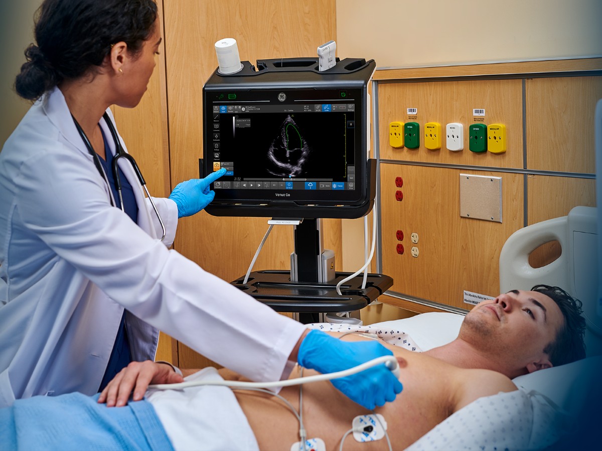
With Real-Time Ejection Fraction (EF), physicians can use AI in ultrasound to quickly and accurately assess patients showing symptoms of heart failure. This semiautomated tool integrates AI in ultrasound to easily calculate the EF of the left ventricular (LV) from only the apical four-chamber (A4CH) view. It also has a built-in color indicator that identifies the quality of the image using red, yellow, or green based on a combination of scan quality, identification of the A4CH view, and consistency of the EF results.
This Real-Time EF tool traces the walls of the LV in each frame and identifies the end-diastolic and end-systolic frames, which can easily be navigated between the acquired heart cycles and end-diastolic and end-systolic frames in the last four seconds, once saved by the user. Another time-saving aspect to this tool is that ECG is not required in order to take an accurate EF measurement. The novel semiautomated Real-Time EF tool helps physicians gain an EF value within three seconds of live scanning in A4CH view, when image quality is sufficient as determined by the Quality Indicator. In 86% of cases, the ejection fraction results from the Real-Time EF have been found to be within ±10 points of experts' measurements.1
As emergency and critical care departments face ongoing crowding concerns, having a fast and accurate tool can elevate the standard of care patients receive and reduce the time it takes to reach consistent EF results.
The Real-Time EF tool could affect the clinical outcomes of critically ill patients, reducing hospital stays and lowering complication rates by replacing time-consuming manual measurements. The GE Venue Family brings this technology to the forefront to provide ease of use and improve care for patients.
Download the whitepaper here.
References:
- Venue and Venue Go R3 technical claims document (DOC2391130). Venue Fit technical claims document (DOC2454794).

