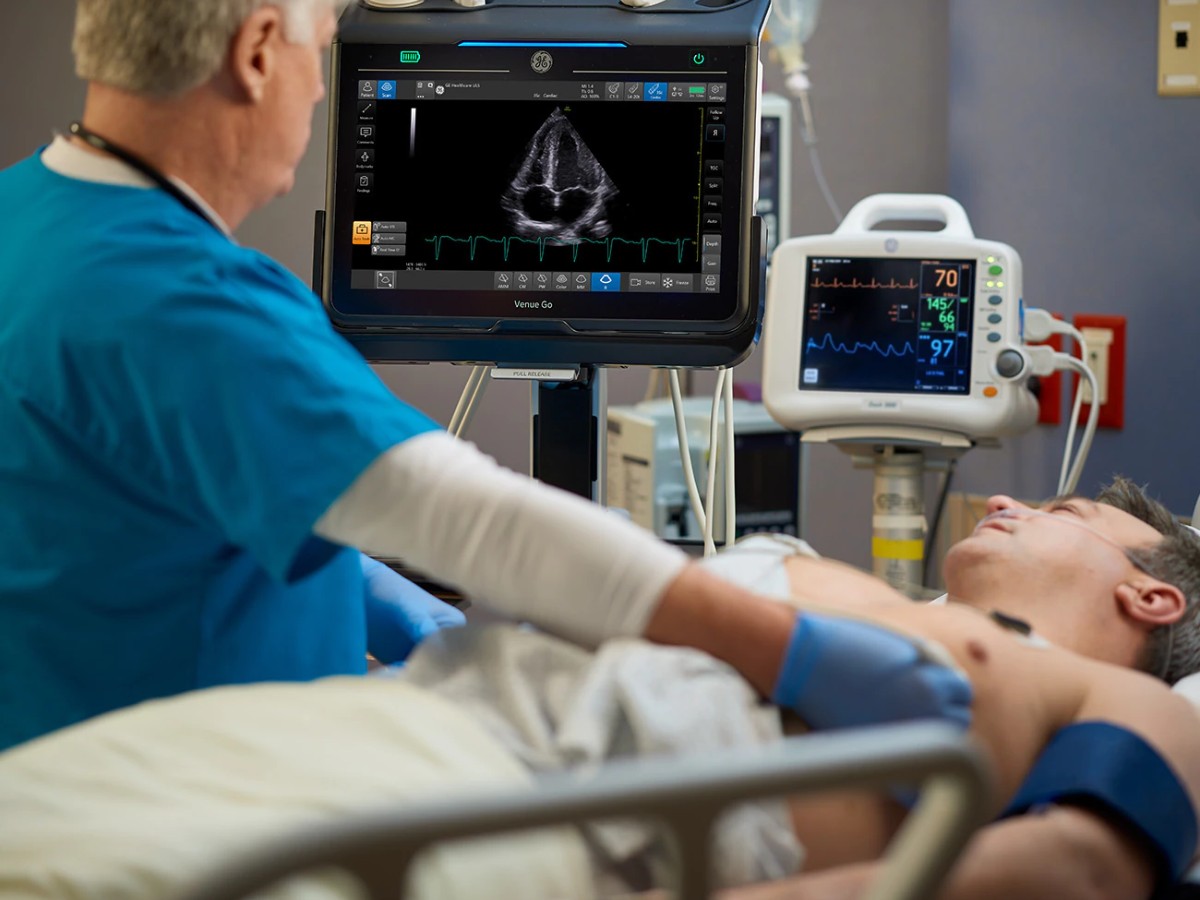
Hospital physicians have long relied on ultrasound for critical diagnostic insights. But for most of that time, the technology was large and costly, meaning it was mostly confined to radiology labs and cardiology and obstetrics units.
Point of Care Ultrasound (POCUS) changed that paradigm. POCUS devices are more portable, less expensive, and easier to use—and they've rapidly become part of the everyday tool kit for clinicians across specialties.
POCUS doesn't replace what traditional ultrasonography does. Both POCUS and traditional ultrasound bring their own benefits, best practices, and use cases.
What Is Point of Care Ultrasound?
As the name suggests, POCUS enters the picture at the point of care—whether that's a hospital bed, a physician's office, or even the sidelines of a sporting event. POCUS systems can include handheld devices that stream real-time sonographic images to phones and tablets as well as portable systems on rolling carts like the GE Venue series.
These portable ultrasound machines provide crisp, detailed images that can provide real-time guidance during a procedure or offer rapid insights that help clinicians make informed decisions quickly.
This makes the technology useful across a variety of medical specialties. It has become an essential tool both in facilities such as emergency rooms and ICUs as well as among roving specialists like anesthesiologists and orthopedists.
Key Differences Between POCUS and Traditional Ultrasound
POCUS differs from traditional ultrasonography, in which comprehensive studies are performed by diagnostic medical sonographers and later interpreted by radiologists. Instead, clinicians perform POCUS scans at the bedside in an attempt to answer specific questions that can help confirm or rule out a suspected diagnosis, narrow the differential, or guide a procedure.1 This information becomes part of the differential alongside data gathered from physical exams, patient histories, and traditional procedures and tests.
Aside from the form factor, there are several key differences between POCUS and regular ultrasound:
Comprehensiveness. Traditional ultrasound assesses an anatomical region using predefined parameters and measurements to provide a diagnosis. POCUS assesses one part of the body at a time to answer very specific questions in the context of a physical exam and patient history—for example, does this patient with lower back pain and fever have kidney stones? Will this patient's airway make intubation difficult?
Expertise. Traditional ultrasonography is complex and nuanced, and images are typically interpreted by radiologists or cardiologists with significant ultrasound experience. POCUS is more user-friendly and can be performed by any POCUS-trained clinician.
Time to results. Depending on radiology's backlog and the urgency of a patient's condition, traditional ultrasound results can take hours or days to receive. With POCUS, clinicians get clinically useful information in real time and can use that information to increase the accuracy of their bedside assessment.
Location. For a traditional ultrasound, the patient must travel to the machine. POCUS, on the other hand, travels to the patient.
Simply put, while a regular ultrasound is a comprehensive diagnostic imaging exam, POCUS acts as one part of a bedside assessment. Although its role is different, it can yield valuable information for clinicians throughout hospital systems.
Use Cases for Point of Care Ultrasound
Across specialties, POCUS-equipped clinicians in emergency, critical care, MSK, and anesthesia settings use the technology for:
Lung Ultrasound
POCUS can help emergency room and ICU doctors detect signs of acute respiratory failure, often secondary to pneumonia, bronchitis, or asthma exacerbation.2 It can also help assess patients for pneumothorax, pulmonary edema, interstitial syndrome, pleural effusion, and lung consolidation.
Cardiac and Vascular Ultrasound
Cardiac ultrasound reveals meaningful information about a patient's cardiac output, fluid status, and valvular pathology.3 It can detect signs of hypovolemic shock, pericardial effusion, pulmonary embolism, cardiac tamponade, and other serious conditions that require immediate attention. Vascular ultrasound of the lower extremities can also help detect deep vein thrombosis, which can lead to pulmonary embolism if left untreated.
Abdominal Ultrasound
Using POCUS to assess a patient's kidneys or liver, clinicians can detect abnormalities that suggest chronic disease or acute injury. Clinicians can also rely on abdominal ultrasonography to screen patients for aneurysm and aortic dissection.
Procedural Ultrasound
POCUS can provide real-time guidance during invasive procedures, helping clinicians visualize venous access, difficult airways, and nerve systems. For example, POCUS may come into use as anesthesiologists perform ultrasound-guided nerve blocks, ER doctors intubate patients with difficult airways, and ICU doctors place central venous catheters.
Musculoskeletal Ultrasound
Physicians can use POCUS to visualize soft tissue tears or infection in muscles, tendons, ligaments, and joint spaces. It can also help them detect foreign bodies in these areas.4 Its portability makes it ideal for treating trauma patients in emergency rooms or injured athletes at sporting events.
Benefits of POCUS
As more clinicians integrate POCUS into their medical practices, use cases continue to arise. So too does the body of research describing the benefits of POCUS:
Improved diagnostics. When used alongside physical exams and other traditional diagnostic tools, POCUS may improve the accuracy of diagnoses while reducing the need for supplemental exams and the likelihood of redundant interventions.5
Faster time to treatment. By providing real-time insights, POCUS can help confirm a diagnosis quickly. In turn, this decreases the time it takes to start treatment. These results indicate that POCUS assessment conducted early among patients with dyspnea or chest pain improves diagnostic accuracy and shortens significantly the time to appropriate treatment.6
Safer procedures. Real-time guidance during invasive procedures decreases the chance of complications. For example, ultrasound-guided regional anesthesia increases the probability of successful nerve block while cutting needle placement time and decrease adverse effects (vascular puncture and local anesthetic systemic toxicity).7 The Agency for Health Research and Quality listed ultrasound-guided vascular access as one of the 12 most effective ways to prevent medical errors.2
Safe monitoring. Ultrasound uses nonionizing radiation, so it can be safely repeated again and again without posing risks to patients. This makes it ideal for monitoring disease progression or recovering injuries.
These features can support clinicians across specialties as they address a range of patient needs.
The Future of Point of Care Ultrasound
The use cases for POCUS continue to evolve as practitioners, governing boards, and specialty organizations recognize the advantages of equipping clinicians with portable ultrasound devices. Over the past few years, both the American College of Physicians and the Society of Hospital Medicine have formally acknowledged the value of POCUS for improving diagnostic speed and patient outcomes.8 The Accreditation Council for Graduate Medical Education has deemed POCUS a core requirement for emergency medicine, and more than half of US-based medical schools have implemented ultrasound training into their internal medicine curriculums.1 Meanwhile, the American Society of Anesthesiologists, the Society of Critical Care Medicine, and other subspeciality organizations have created best practices and learning resources to help practicing physicians use POCUS effectively.
Between the diverse utility and expanded access to training, POCUS has earned a place as a reliable bedside tool. The global POCUS market is predicted to grow by a 5-year compound annual growth (CAGR) of nearly 5%.9 POCUS won't replace traditional ultrasound anytime soon—if ever. Nonetheless, it will continue to evolve and prove its worth in hospitals as more clinicians learn how to make the most of this technology.
References
- Koratala A and Reisinger N. POCUS for nephrologists: basic principles and a general approach. Kidney360. 2021;2(10):1660-1668. 10.34067/KID.0002482021.
- Burton L, Bhargava V, and Kong M. Point-of-care ultrasound in the pediatric intensive care unit. Frontiers in Pediatrics. 2022;9. 10.3389/fped.2021.830160.
- Levitov A, Frankel H, Blaivas M, et al. Guidelines for the appropriate use of bedside general and cardiac ultrasonography in the evaluation of critically ill patients—part II: cardiac ultrasonography. Critical Care Medicine. 2016;44(6):1206-1227. 10.1097/CCM.0000000000001847.
- Chen KC, Chor-Ming A, Chong CF, et al. An overview of point-of-care ultrasound for soft tissue and musculoskeletal applications in the emergency department. Journal of Intensive Care. 2016;4:55. https://doi.org/10.1186/s40560-016-0173-0.
- Zieleskiewicz L, Lopez A, Hraiech S, et al. Bedside POCUS during ward emergencies is associated with improved diagnosis and outcome: an observational, prospective, controlled study. Critical Care. 2022;25(1):34. 10.1186/s13054-021-03466-z.
- Golan Y, Sadeh R, Mizrakli Y, et al. Early point-of-care ultrasound assessment for medical patients reduces time to appropriate treatment. Ultrasound in Medicine and Biology. 2020;46(8):1908-1915. 10.1016/j.ultrasmedbio.2020.03.023.
- Muacevic A and Adler JR. Perioperative point-of-care ultrasound use by anesthesiologists. Cureus. 2021;13(5):e15217. 10.7759%2Fcureus.15217.
- Wong J, Montague S, Wallace P, et al. Barriers to learning and using point-of- care ultrasound: a survey of practicing internists in six North American institutions. Ultrasound Journal. 2020;12:19. 10.1186/s13089-020-00167-6.
- Harris, S. Ultrasound equipment – world market – 2019. Signify Research. https://signifyresearch.s3.eu-west- 2.amazonaws.com/app/uploads/2018/06/01114306/B-2019ULS- Ultrasound-Equipment-2019-01.08.19.pdf. May 2019.

