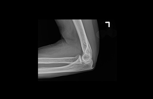
X-ray imaging is a crucial diagnostic tool used in medical facilities worldwide. It is often the first imaging modality used to visualize the internal structures of the body and can provide valuable information for diagnosis and treatment planning. Radiologists play a crucial role in interpreting these images and providing accurate diagnoses, making the quality of the images essential for effective patient care. New solutions in X-ray image processing have emerged as powerful tools enabling image presentation consistency and aiding in the interpretation of complex images.
Artificial intelligence solutions in X-ray
In addition to advances in X-ray detector technology, the implementation of artificial intelligence (AI) solutions in X-ray is revolutionizing advanced image processing. AI tools are playing a crucial role in X-ray, enabling radiologists to process extensive amounts of imaging data through advanced X-ray image reconstruction algorithms. Sophisticated AI solutions, such as deep learning algorithms, can further aid image reconstruction by enhancing the consistency and accuracy of imaging data. AI can be used to improve the consistency of brightness and contrast from image to image, which can make images easier to interpret. Deep learning algorithms can be trained to process and present images differently for specific anatomies, which supports radiologists’ ability to make more accurate diagnoses.
Industry partners such as GE HealthCare are committed to facilitating radiologists’ access to consistent, high-quality X-ray images in radiology to inform diagnoses and improve health outcomes.
“GE HealthCare is focused on facilitating access to the highest quality X-ray imaging possible for our customers,” said Laura Hernandez, Chief Marketing Officer for Women’s Health and X-ray, GE HealthCare. “We’re harnessing the power of our X-ray detectors and developing robust AI tools and image processing solutions that can enhance X-ray images quickly and efficiently so that radiologists have the finely detailed images they need to make accurate and timely diagnoses for their patients.”
AI and automation for X-ray image consistency
Automating tasks during the imaging acquisition process and standardizing imaging protocols can improve image quality and decrease variability in imaging. Reducing inconsistency and variability in X-ray imaging with comprehensive solutions such as on-device automations, ergonomic features, and sophisticated AI algorithms can help ensure that radiologists consistently receive high-quality X-ray images for interpretation.
On-device automations to reduce X-ray scatter are used to adjust how X-ray images are presented to the reading radiologist. A variability that is not part of a desired signal, image noise can be caused by a number of factors, including scatter, and can make it difficult to see important details in X-ray images, which can lead to images that aren’t of diagnostic quality.
Traditionally, X-ray technologists place an anti-scatter grid in front of the detector to remove scatter X-ray photons that do not otherwise contribute to image quality. An on-device software solution from GE HealthCare can replicate the contrast enhancement of imaging with a physical anti-scatter grid, eliminating the extra step in the workflow of the X-ray technologist.
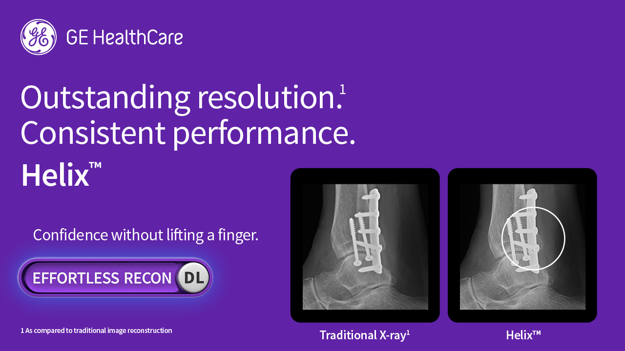
Advanced image processing solutions can also be used to sharpen images, making them clearer and easier to interpret. GE HealthCare’s advanced AI-driven solution for X-ray image processing allows to customize the brightness and contrast levels for 58 anatomy / view combinations while maintaining the full resolution of the images from the X-ray detector. And because the original image data is not altered by the AI, the radiologist maintains the freedom to quickly and easily adjust brightness and contrast as needed.
Assistance from sophisticated image processing and AI solutions can help to reduce the time it takes to interpret X-rays. With the automation of many of the tasks involved in image interpretation, radiologists can focus on more complex tasks, such as making diagnoses and developing treatment plans.[1]
Deep learning for X-ray
Increasingly used across radiology, deep learning algorithms are also having a major impact in X-ray, such as using AI to identify patterns that are associated with certain diseases, such as lung nodules or other abnormalities.
Using deep learning to help generate X-ray image display properties can help radiologists by adjusting image settings. Onsite, each user can set their preferred settings to enable optimal image presentation to the radiologist. For example, the fine detail needed for an X-ray image of the hand is much different from that of a chest X-ray image.
A bright future for image quality in X-ray
The use of AI tools and advanced image processing to support X-ray image consistency and quality is a promising development that has the potential to improve accuracy and efficiency in radiology. As AI technology continues to develop, it is likely that there will be even more innovative applications of AI across radiology in the years to come. GE HealthCare is committed to providing powerful, AI-driven solutions that deliver continued improvements in X-ray image quality and across radiology.
RELATED CONTENT
Learn more about GE HealthCare’s X-ray solutions, here.
Learn more about GE Healthcare’s X-ray Helix™ Advanced Imaging Processing
DISCLAIMER
Not all products or features are available in all geographies. Check with your local GE HealthCare representative for availability in your country.
REFERENCE
Improving presentation consistency of radiographic images using deep learning | (2021) | Maheen Aboobacker | Publications | Spie