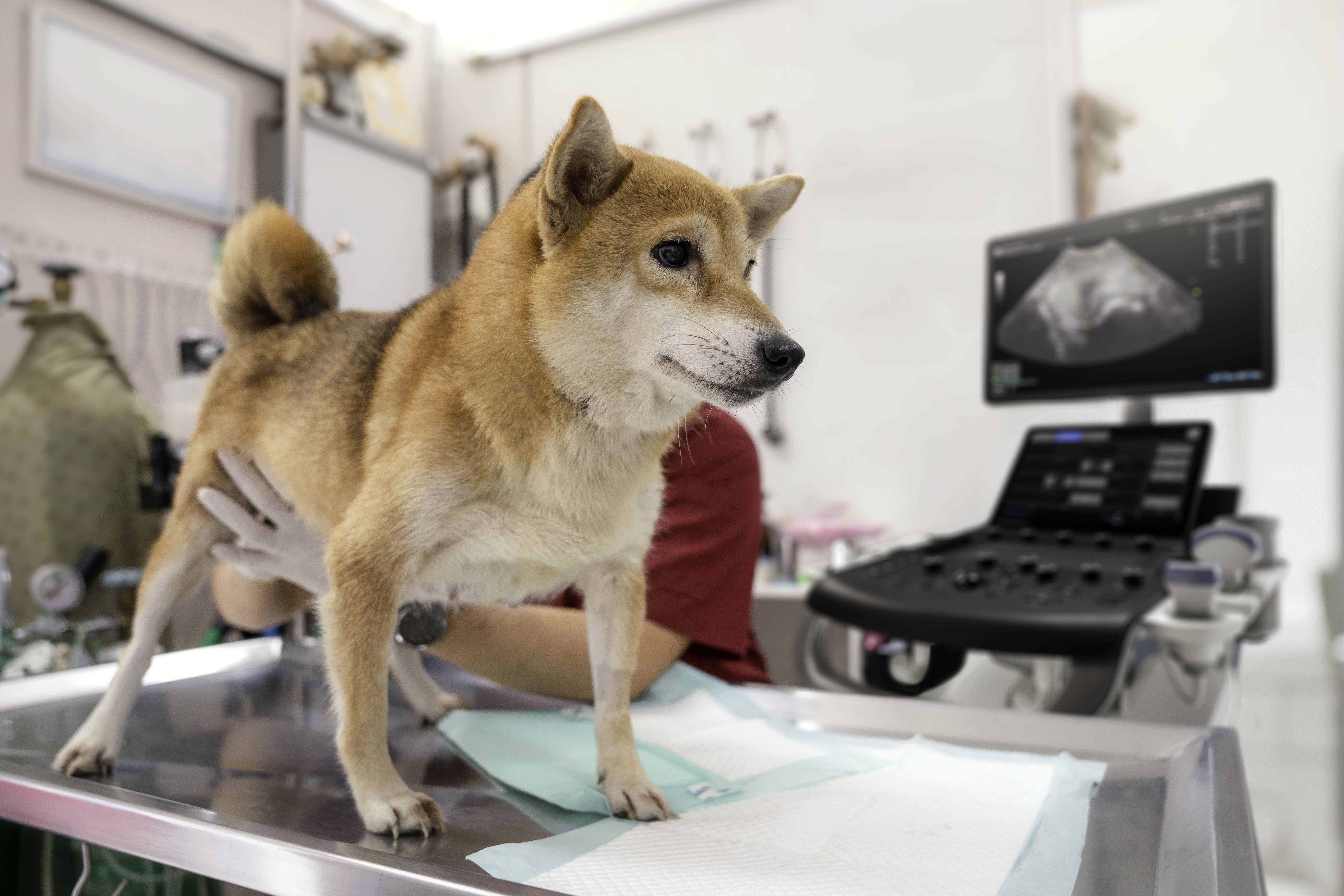
Veterinary medicine is experiencing a rapid increase in ultrasound utilization across virtually all types of practice, from emergency to primary care. Recent data from the University of North Carolina indicate that more than half of all veterinary practices currently have some kind of veterinary ultrasound system, with 45% performing at least five exams every week.1
While abdominal and thoracic imaging are among the most common applications for ultrasound in veterinary practices,2 veterinarians at all experience levels and specialties are using this technology to diagnose and manage a wide range of conditions.
This article examines the clinical and operational reasons for this proliferation, the benefits to your practice, and factors to consider when choosing or upgrading your system.
Patient-focused benefits of in-office veterinary ultrasound
Let's start with the most basic premise: veterinary medicine differs from traditional clinical practice in that animals can't articulate their problems or concerns. Veterinarians often spend valuable time trying to deduce the timeline of symptom onset and any causative correlation based on what the animal may have been doing.
Meanwhile, external ultrasound referrals can take weeks, or even months, particularly if the danger does not appear to be immediate. During this waiting period, problems can worsen beyond the scope of treatment. In-office ultrasound can significantly expedite the diagnostic process, often leading to lifesaving interventions for critical health issues.
Ultrasound applications in veterinary medicine
Among the many time-sensitive veterinary health conditions that ultrasound has been able to detect are:
- Cardiac issues. In emergency care environments, cardiac ultrasounds can examine the heart and its surrounding structures, including the pericardial sac.3 Integrating ultrasound into your practice helps your patients avoid unnecessary trips to the emergency clinics, allowing quick determination of the location, scope, and nature of their animals' cardiac event and the presence of internal bleeding, pneumothorax, or other issues. The application of focused ultrasound has been demonstrated to improve the accuracy of cardiac diagnosis, specifically in feline heart conditions.4
- Liver disease. Ultrasound has proven effective in detecting abnormally fatty livers in dogs,5 as well as lesions, growths, and other abnormalities that can indicate more severe health problems.6 Having adequate diagnostic resources at your disposal will help support your practice to identify or rule out different types of hepatic disease.
- Thyroid issues. Ultrasound can help identify feline hypothyroidism,7 as well as a wide range of other thyroid conditions in veterinary care.8 It can also help detect nodules that indicate hyperparathyroidism in dogs.9
- Malignant canine mammary tumors. Tumors of the breast are among the most common types of growth experienced by companion animals, specifically female dogs. Breast ultrasound is an effective and noninvasive way of evaluating tissue architecture, allowing for optimal imaging of small structures. The superficial positioning of the mammary glands allows transducers to get the highest-quality scans for early detection and evaluation.10
- Adrenal gland issues. Focused ultrasound in primary care veterinary medicine can help to identify adrenal tumors, glandular lesions, and a range of other potentially serious issues more quickly and accurately.11 Lesions and growth size have been identified as the most accurate ultrasonographic indicators of adrenal disorders.
One of the most common and effective applications of veterinary ultrasound, is in obstetrics, where it's often used to assess, locate, and monitor pregnancies. Other areas of application for veterinary ultrasound include tendons, ligaments, and urinary tract. It's also a natural diagnostic next step upon detection of abnormalities in animals' blood, urine, or stool, and can assist with tissue collection and biopsies.
Diagnosis of these and other condition areas often requires imaging referrals by a specialist. Private-practice veterinarians trained in ultrasound can help to reduce these wait times and develop a care plan more quickly.
Primary care veterinarians in rural areas, in particular, should consider investments in ultrasound. Rather than referring animals to hospitals or specialty clinics, they can expedite treatment by diagnosing at the time of care.
Revenue and growth benefits of in-office veterinary ultrasound
Each of the above condition areas represents an opportunity for veterinarians who integrate primary care ultrasound to grow their practices while also helping their patients. Apart from aiding clinical decision-making, ultrasound scans also represent an additional revenue opportunity for the veterinary practice.
Offering ultrasound at your clinic can also distinguish your practice as a center of diagnostic excellence in specific condition areas and help you keep elements of treatment under your roof for continuity of care. In the next section, we examine how the right system can help you hit the ground running, flatten the learning curve, and let you and your staff start reaping the growth benefits of ultrasound immediately.
Certain veterinary ultrasound vendors offer flexible financing agreements so you don't expend all of your procurement dollars at once. At the same time, they provide comprehensive warranty agreements and robust training, building peace of mind and maximum assurance that clinicians of all experience levels can get the most out of their system. This support is particularly important for clinics with limited budgets and resources.
Factors to consider when choosing a veterinary ultrasound system
When choosing an ultrasound for your veterinary practice, whether it's your first system or you're looking to upgrade, there are several core elements to consider.
- Diversity of clinical application. Veterinarians must assess an incredibly broad range of anatomies every day. For that reason, it's essential to choose a system that can seamlessly perform a wide array of scans effectively and efficiently through the use of different probes and presets. The ability to scan cardiac, thyroid, vascular, abdominal, musculoskeletal, obstetrics, and more with one system is essential.
- Easy scalability. Your system should also provide opportunities to upgrade with minimal disruption to your daily operations. With a range of transducers and applications that enhance the amount of information you see during patient exams; your practice can expand by adding probes and available software.
- Enhanced technology for image optimization and scan performance. Today's systems have more features than ever to maximize image quality and scan accuracy. Needle recognition simplifies ultrasound-guided biopsies or injections by continuously clarifying point positioning. Some veterinary ultrasound systems have features that continuously optimize brightness, contrast, gain, and uniformity of images while scanning different tissues. Other features, like body markers and annotations, can quickly label images. 3D/4D capability can guide decision-making by enabling additional dimensions in real-time. Spatial recognition features such as elastography can also show the distribution of tissue elasticity properties in a region of interest, by estimating the strain before and after tissue distortion.
- Improved workflow and reliability. Some veterinary practices may lack sufficient training and personnel capacity to effectively integrate ultrasound into their daily routine. In response, certain systems offer a simplified workflow and animal-specific report templates, with the follow-up tools to support new users and understaffed practices to perform the most accurate scans.
Images that speak for your patients
Veterinarians in the primary care landscape have a unique opportunity to improve patient care while growing their practice. With more veterinarians investing in this game-changing diagnostic resource each month, consider whether ultrasound may be the next stage of evolution for your practice.
Resources:
-
DeFrancesco T, Royal K. A survey of point-of-care ultrasound use in veterinary general practice. Education in the Health Professions. 2018;1(2): 50. https://doi.org/10.4103/ehp.ehp_21_18.
-
Pelchat J, Chalhoub S, Boyson SR. The use of veterinary point-of-care ultrasound by veterinarians: A nationwide Canadian survey. Canadian Veterinary Journal. December 2020. https://www.ncbi.nlm.nih.gov/pmc/articles/PMC7659883/.
-
Lyssens A, Lekane M, Gommeren K, et al. Focused cardiac ultrasound to detect pre-capillary pulmonary hypertension. Frontiers in Veterinary Science. 2022; 9. https://doi.org/10.3389/fvets.2022.830275.
-
Janson CO, Hezzell MJ, Oyama MA, et al. Focused cardiac ultrasound and point‐of‐care NT‐proBNP assay in the emergency room for differentiation of cardiac and noncardiac causes of respiratory distress in cats. Journal of Veterinary Emergency and Critical Care. 2020; 30(4): 376–383. https://doi.org/10.1111/vec.12957.
-
Assawarachan SN, Chuchalermporn P, Maneesaay P, et al. Evaluation of hepatobiliary ultrasound scores in healthy dogs and dogs with liver diseases. Veterinary World. 2019; 12(8): 1266–1272. https://doi.org/10.14202/vetworld.2019.1266-1272.
-
Kumar V, Kumar A, Varshney AC, et al. Diagnostic imaging of canine hepatobiliary affections: a review. Veterinary Medicine International. 2012. https://doi.org/10.1155/2012/672107.
-
Nussbaum LK, Scavelli TD, Scavelli DM, et al. Abdominal ultrasound examination findings in 534 hyperthyroid cats referred for radioiodine treatment between 2007-2010. Journal of Veterinary Internal Medicine. 2015; 29(4): 1069–1073. https://doi.org/10.1111/jvim.13369.
-
O'Neill DG, Khoo JSP, Brodbelt DC, et al. Frequency, breed predispositions and other demographic risk factors for diagnosis of hypothyroidism in dogs under primary veterinary care in the UK. Canine Medicine and Genetics. 2022; 9(1). https://doi.org/10.1186/s40575-022-00123-8.
-
Pollard RE, Bohannon LK, Feldman EC. Prevalence of incidental thyroid nodules in ultrasound studies of dogs with hypercalcemia (2008-2013). Veterinary Radiology & Ultrasound. 2014; 56(1): 63–67. https://doi.org/10.1111/vru.12181.
-
Feliciano MAR, Uscategui RAR, Maronezi MC, et al. Ultrasonography methods for predicting malignancy in canine mammary tumors. PloS One. 2017; 12(5): e0178143. https://doi.org/10.1371/journal.pone.0178143.
-
Pagani E, Tursi M, Lorenzi C, et al. Ultrasonographic features of adrenal gland lesions in dogs can aid in diagnosis. BMC Veterinary Research. 2016; 12(1): 267. https://doi.org/10.1186/s12917-016-0895-1.

