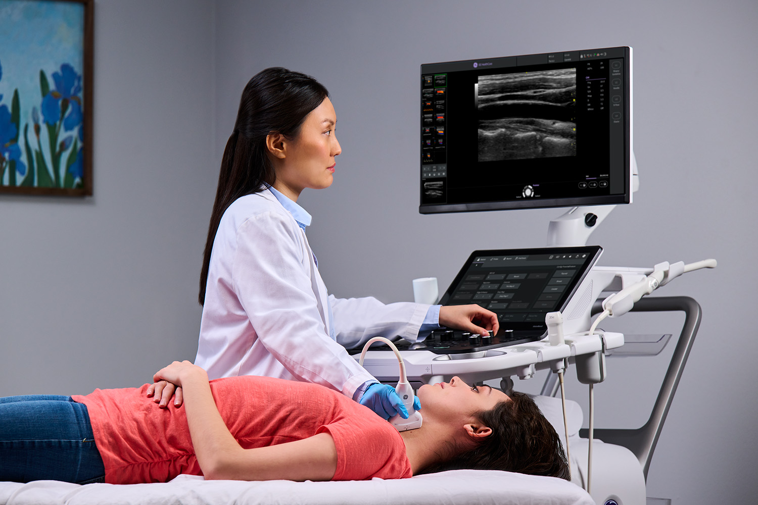
The advanced treatment of vascular disease has historically been the purview of vein specialists:1 while the process may start in general practitioners' offices, they typically refer patients to a specialist for the next stage of treatment. In the past, this process was necessary to optimize care and ensure patients received the most informed guidance possible. However, the advent of in-office vascular ultrasound is changing the game, allowing for the early detection of manageable, preventable diseases that affect millions of people globally each year.
Thanks to rapid and significant recent innovations, a growing number of general practitioners can now provide vascular ultrasound in their offices. This technology increases diagnostic confidence, accelerates treatment, and improves the overall patient experience across a wide range of vascular conditions. Clinicians can also utilize vascular ultrasound to guide interventions for a number of mild, moderate, and severe vein conditions that general practitioners see daily.
What are ultrasound-guided vascular interventions, and what conditions can they treat?
Ultrasound-guided vascular interventions have made peripheral access considerably easier in recent years. In the past, individual anatomy, co-occurring conditions that can inhibit vein access, operators' skill levels, and other factors have made vascular intervention more difficult, resulting in higher rates of failure and complication.2
High-resolution imaging enables clinicians to identify pertinent vascular health issues while reducing the number of access attempts through other more invasive measures.
When properly applied, ultrasound guidance for vascular access improves success rates while reducing iatrogenic injury, the number of needle passes, and infection rates.3 It can also help to improve the overall patient experience by increasing comfort during treatment.4
Proactive intervention: Using ultrasound to detect broader vascular-based health issues
Many significant health issues start in the blood vessels; non-invasive vascular imaging can help identify and treat such issues more quickly. For example, tell-tale vascular symptoms, such as varicose or spider veins can indicate venous insufficiency. Providers can also more easily identify complications like coagulation, atherosclerosis, valvular incompetence, peripheral vascular disease, and many others through primary care ultrasound.
While it can play a key role in preventive medicine, ultrasound-guided vascular intervention is largely underutilized in primary care environments. Healthcare providers can apply vascular ultrasound to identify and treat disease in the following ways:
-
Checking for atherosclerosis. General practitioners can use a simple, non-invasive vascular ultrasound to detect narrowing of the carotid artery and determine the best course of action for at-risk patients. This condition, caused by a build-up of plaque, can significantly increase the risk of stroke.5 Clinicians may recommend pharmacological intervention, vascular surgery, or simple lifestyle changes, such as diet adjustments, smoking cessation, stress management, and increased exercise.
-
Identifying peripheral artery disease (PAD). Peripheral artery disease, a common vascular condition affecting the legs and feet, can cause significant circulatory problems in the lower extremities and decrease patients' quality of life. Using vascular ultrasound allows general practitioners to assess claudication, rest pain, and skin changes that can indicate PAD6, and identify the extent of stenosis and determine the best course of treatment.
-
Managing type 2 diabetes complications. Vascular ultrasound can help identify restricted blood flow to the lower extremities, which has critical clinical significance for the evaluation and monitoring of diabetes. Considering the wide gap in diabetes education and support, ultrasound can help clinicians educate patients on the seriousness of their conditions, motivating self-management under the supervision of their trusted primary care physician.
Facilitating comfort and collaboration between GPs and patients
The bottom line is ultrasound is a non-invasive, comfortable, convenient, and accurate way to identify vascular issues that can signify the onset and progression of a wide range of vascular conditions. It can help clinicians inform and empower patients, helping them to become more active in their own care, whether their conditions result from genetic predispositions, the natural aging process, or lifestyle issues.
In a preventive setting, vascular ultrasound can greatly lower the risk of progression for many conditions. In terms of immediate treatment, it can illuminate a faster and more direct route to care, whether through emergency surgical intervention or the creation of an ongoing treatment plan.
Most patients prefer a centralized care environment, with as many diagnostics and interventions as possible offered in one place. Beyond expediting diagnosis and treatment, centralized care enables faster and more direct answers from their primary care physicians.
In some areas of the world, an ultrasound referral can take weeks or even months.7 By investing in vascular imaging capabilities and integrating them into preventative treatment plans, general practitioners can help their patients avoid these wait times, which can reduce quality of life and increase morbidity.
It's also important, however, to choose the ultrasound system that best suits your practice needs, resources, and growth objectives.
What to look for in a system for vascular ultrasound
Whether you're upgrading your system or making a first-time investment, current options make it easier than ever for users of all experience levels to obtain vascular imaging in the office. Look for a system that offers:
-
The latest image-optimization features to help you achieve the most accurate real-time scans
-
Workflow features such as virtual convex to enable a larger field of view or follow up tools to allow for real-time comparison of past and present images, helping to assess the progression of vascular disease
-
Measurement packages for fast, easy, and accurate automatic velocity measurements and carotid intima-media-thickness (IMT) to access atherosclerosis and cardiovascular disease risk
-
Portability and an ergonomic design that adapts to all environments, no matter the size or configuration of space
-
Systems which offer intuitive and dynamic low blood flow imaging with power Doppler imaging (PDI), B-Flow and B-Flow Color
-
The ability to easily upgrade through remote software installations and probe additions
The primary barriers to ultrasound integration in any area of specialization for general practitioners include a steep learning curve and high costs. However, the right system should alleviate these concerns, with comprehensive training and education and flexible financing options that allow a minimal one-time investment.
Finally, in addition to improving patient care and health outcomes, integrating vascular ultrasound into your practice can create an entirely new revenue stream in a very short time. In short, vascular ultrasound in primary care is beneficial for patients and providers alike.
Learn more about primary care ultrasound and how it can transform patient care through vascular imaging.
Resources:
-
Young M. Vascular medical quality and patient safety. StatPearls - NCBI Bookshelf. February 2023. https://www.ncbi.nlm.nih.gov/books/NBK578196/
-
Dietrich CF, Horn R, Morf S, et al. Ultrasound-guided central vascular interventions, comments on the European Federation of Societies for Ultrasound in Medicine and Biology guidelines on interventional ultrasound. Journal of Thoracic Disease. 2016; 8(9): E851–E868. https://doi.org/10.21037/jtd.2016.08.49
-
McGee DC, & Gould MK. Preventing complications of central venous catheterization. The New England Journal of Medicine. 2003; 348(12): 1123–1133. https://doi.org/10.1056/nejmra011883
-
AIUM Practice parameter for the use of ultrasound to guide vascular access procedures. Journal of Ultrasound in Medicine. 2019; 38(3). https://doi.org/10.1002/jum.14954
-
Fedak A, Chrzan R, Chukwu O, et al. Ultrasound methods of imaging atherosclerotic plaque in carotid arteries: examinations using contrast agents. Journal of Ultrasonography. 2020; 20(82): e191–e200. https://doi.org/10.15557/JoU.2020.0032
-
Nasra K. Sonography vascular peripheral arterial assessment, protocols, and interpretation. StatPearls - NCBI Bookshelf. March 2023. https://www.ncbi.nlm.nih.gov/books/NBK570577/
-
Imperial College Healthcare. Patient information. Imperial College Healthcare NHS Trust. August 2023. https://www.imperial.nhs.uk/our-services/surgery/vascular-surgery/patient-information
JB31599XX

