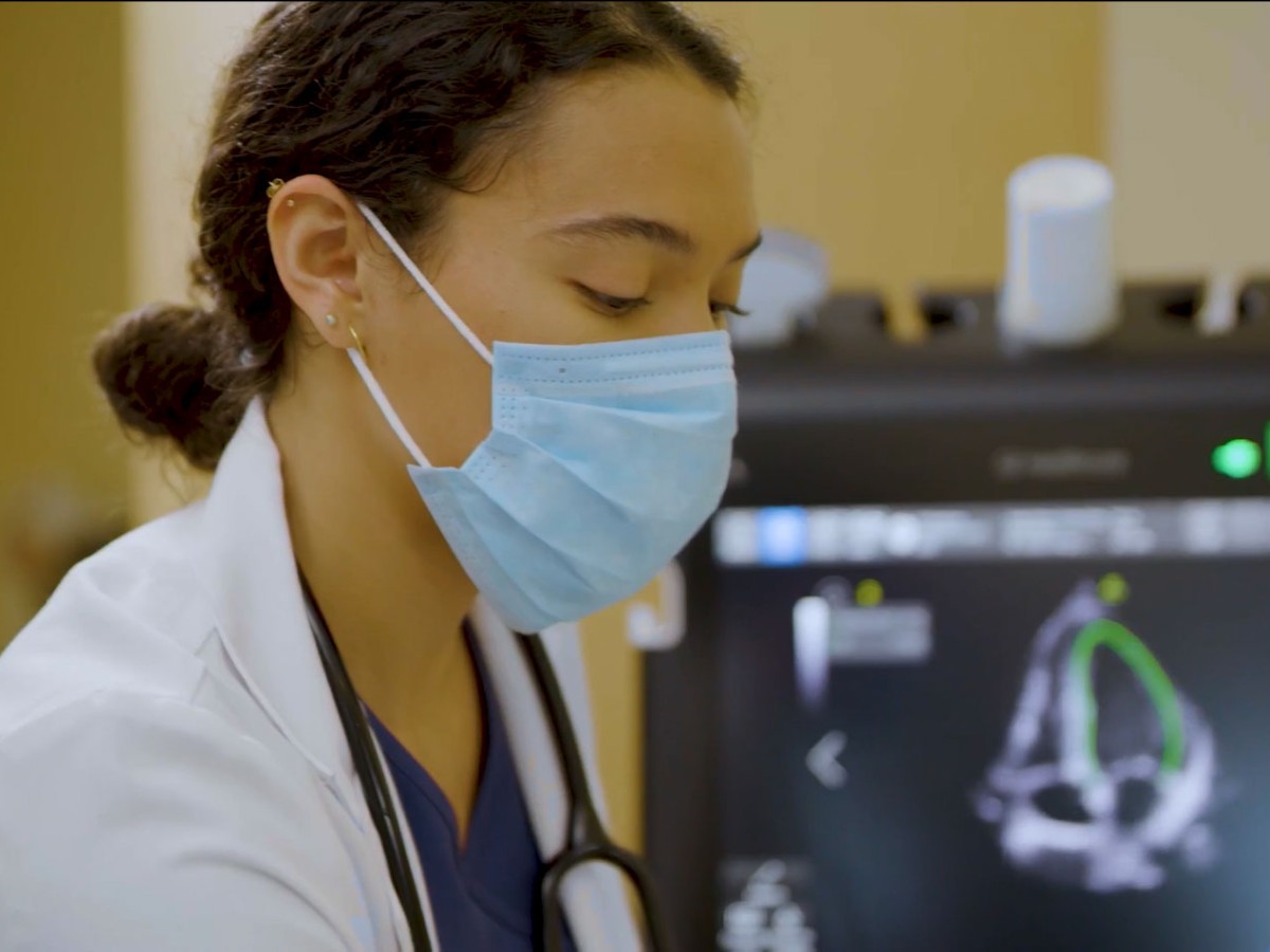
The Point of Care Ultrasound (POCUS) exam has revolutionized health care, encouraging physicians to tackle challenges their counterparts 25 years ago could not have envisioned.
A POCUS exam can help care teams meet growing demands as they address more serious cases and keep up with a higher number of patients in both hospital and clinic settings. Physicians can practice cost-effective medicine while providing an accurate and time-sensitive evaluation.
Examination via POCUS has clear benefits for practicing physicians—and, in turn, for the ones under their care.
How the POCUS Exam Benefits Physicians
The wide-reaching benefits of the POCUS exam have earned it a place in both inpatient and outpatient settings. When used in conjunction with a patient's history, physical examination, and lab results, POCUS offers valuable information to help guide a physician's clinical decision-making. Additionally, POCUS goes to where the patient is and is performed by the physician rather than an ultrasound technician; this not only decreases the need to transport patients for additional testing but also boosts patient-physician interaction time. In many instances, POCUS can complement advanced imaging and be used to determine if more invasive procedures are required, which has widespread benefits including cost savings and patient safety.
The Role of POCUS in the Emergency Department
POCUS has extensive uses throughout the emergency department (ED). The use of POCUS in the ED started with the focused assessment with sonography for trauma (FAST) exam and has expanded widely since then. Physicians in the ED practice under constant pressure to see, evaluate, and treat patients quickly. Access to POCUS exam allows them to do so.
Evaluating Respiratory Distress
Respiratory distress is a common complaint and with a wide differential diagnosis. A clinical exam can be helpful in diagnosing respiratory distress, but imaging to augment the exam findings further narrows the differential. POCUS of the lung shows 94% sensitivity in assessing pleural effusion and 92% sensitivity for pulmonary contusion.1 Furthermore, using POCUS as compared to the standard of care (i.e., chest X-ray or CT scan) reveals a 4.5 hour decrease in time to accurate diagnosis for these patients.2. As POCUS becomes a part of the clinical equipment of every ED physician, it has the potential to alter the standard of care and vastly improve ED wait and transfer times.
Monitoring Small Bowel Obstruction
Two percent to 4% of patients who present to the ED with abdominal pain are diagnosed with small bowel obstruction (SBO).3 Unfortunately, SBO is hard to diagnose based on physical exams and lab tests, necessitating imaging of some sort for further assessment. These patients are typically directed to undergo a CT scan, which can be expensive and time-consuming.
POCUS is 92.4% sensitive and 96.6% specific for screening symptoms for SBO, which is similar to a CT scan.3 Based on projection models, for patients ultimately diagnosed with SBO, opting to use POCUS instead of a CT scan could save around $30 million annually in health care costs.3 Furthermore, because POCUS can be performed at the bedside, it allows for faster screening and definitive discharge of the patient to the floor or ambulatory setting.
POCUS in the Inpatient Setting
Beyond the ED, POCUS is used in many other inpatient hospital settings.
Using POCUS in Critical Care
Physicians who take care of patients in the ICU stand to benefit from POCUS. Critically ill patients may have rapidly changing clinical statuses that require quick assessment and decision-making. Unstable patients can also present challenges during transport. POCUS allows critical care physicians to evaluate these patients rapidly at the bedside.
Using POCUS, a critical care physician can scan a patient's heart, inferior vena cava, lungs, and abdomen—all within six minutes at the bedside.5 Combining these results with findings from a physical exam and lab is a path to more precise diagnostics.5
Detecting Deep Vein Thrombosis
Patients in the hospital are at comparatively high risk of deep vein thrombosis (DVT). POCUS offers a sensitive and specific method of evaluation: POCUS exams of femoral and popliteal veins can have a 96% sensitivity and 97% specificity for detecting DVT.1 Furthermore, this exam is easy to learn; after just two hours of training, physicians found DVT with 90% sensitivity and 97% specificity.1 With repeat examinations, diagnostic accuracy can approach 100%.1
POCUS in the Outpatient Setting
In the outpatient setting, POCUS helps physicians improve access to timely care, raise patient satisfaction, and reduce the need for costly tests.
Improving Access to Care
Office-based physicians don't always face the same time pressures as their counterparts in the inpatient setting, but they still rely on time-sensitive access to diagnostic exams. Often, this requires scheduling patients for imaging or testing in a different setting from their office and having to follow up with results at another appointment. Utilizing POCUS allows physicians to do the screening in their office and create a care plan in one appointment.
Turning to POCUS for Thyroid Nodules
Endocrinologists are seeing an increasing number of clinic patients with incidental thyroid nodules, inviting a differential diagnosis that includes both benign nodules and malignancy. Although the vast majority of these nodules are benign, they still require a workup so as not to miss the 5% that are malignant.6
If the ultrasound showed malignant features, these patients would traditionally head to radiology for further ultrasound examination and possible biopsy. Benign and malignant thyroid nodules have different characteristics on ultrasound imaging that are relatively straightforward to learn.7 With POCUS, endocrinologists have the ability to identify the nodules in the clinic and determine which patients have malignant features that require biopsy in radiology. Since most thyroid nodules are benign, POCUS can help save the vast majority of patients from further imaging and clinic appointments.
How POCUS Exams Decrease Healthcare Costs
Another advantage of the POCUS examination is its ability to decrease costs. Because POCUS can reduce the need for more expensive imaging modalities and is quick to perform, patients may be discharged faster, especially in the ED. One study found POCUS use eliminated on average $1,000 of additional testing for privately insured patients, nearly $3,000 for out-of-network or uninsured patients, and more than $150 for Center for Medicare and Medicaid Services patients.8 Because insurance plans are moving away from fee-for-service and toward bundled payments, the fewer tests that need to be performed, the more money the hospital system stands to save.
The vast number of POCUS exam benefits creates opportunities for better care in both the inpatient and outpatient settings, making it a critical tool for physicians. Using POCUS allows physicians to practice time-sensitive, cost-effective medicine—ultimately shortening the time to effective treatment for patients in need.
References:
-
Arnold M, Jonas C, Carter R. Point-of-care ultrasonography. American Family Physician. March 1, 2020. 101(5):275-285. https://www.aafp.org/afp/2020/0301/p275.html
-
Gaber H, Mahmoud M, et al. Diagostic accuracy and temporal impact of ultrasound in patients with dyspnea admitted to the emergency department. Clinical and Experimental Emergency Medicine September 2019. 6(3): 226-234. https://doi.org/10.15441/ceem.18.072.
-
Brower C, Baugh C, et al. Point-Of-Care ultrasound-first for the evaluation of small bowel obstruction: national cost savings, length of stay Reduction, and preventable radiation exposure. Academic Emergency Medicine. February 20, 2022. https://doi.org/10.1111/acem.14464.
-
Fabrice D, Grimaldi D, et al. Timing and causes of death in septic shock. Annals of Intensive Care. June 2015; 5(16). https://doi.org/10.1186/s13613-015-0058-8.
-
Melgarejo S, Noble V, Schaub, A, Point of care ultrasound: an overview. American College of Cardiology. Oct 2017. https://www.acc.org/latest-in-cardiology/articles/2017/10/31/09/57/point-of-care-ultrasound.
-
Bernet V, Chindris A. Update on the evaluation of Thyroid Nodules. Journal of Nuclear Medicine. July 2021. 62(2):13S-19S. https://doi.org/10.2967/jnumed.120.246025.
-
Durante C, Grani G. The diagnosis and managment of thyroid nodules: a review. JAMA. March 2018. 319(9):914-924. https://jamanetwork.com/journals/jama/article-abstract/2673975.
-
Van Schaik G, Van Schaik K, Murphy M. Point-of-care ultrasonography (POCUS) in a community emergency department: an analysis of decision making and cost savings associated with POCUS. Journal of Ultrasound Medicine. August 2019. 38(8):2133-2140. https://doi.org/10.1002/jum.14910.

