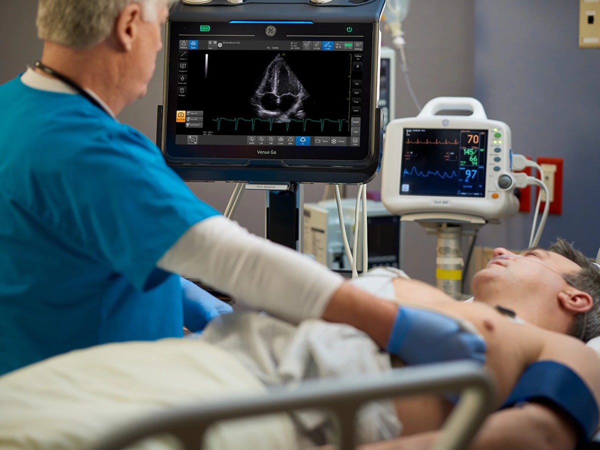
When patients present to the emergency department (ED) with symptoms of heart failure or other cardiac conditions, diagnostic imaging tests are usually a reasonable first step. Many imaging exams require the presence of radiology, but cardiac Point of Care Ultrasound (POCUS) does not—and it is gaining ground in EDs as an option to evaluate for heart failure assessment as well as other heart conditions.
The portability and ease POCUS offers physicians further insight into what's going on within a patient, offering further tools to help triage, guide resuscitation, and determine clinical pathways.
Using POCUS to Examine Heart Conditions
As its convenience and ease of use become more well-known, POCUS has become increasingly widespread as part of ED assessment for cardiac conditions. The American College of Emergency Physicians (ACEP) lists specific applications in the ED for cardiac POCUS, including directing a pathway for people with heart failure and evaluating the cause of shortness of breath.1
Dyspnea
Dyspnea, or difficulty breathing, is a common symptom of heart failure, but it can also signal a range of other health issues. Heart failure is one of the leading causes of hospitalizations,2 and many of these patients are at risk of readmission. So, assessing the cause of dyspnea quickly is essential to triaging patients adequately in the ED.
When patients present with difficulty breathing, their diagnosis may include an echocardiogram and B-type natriuretic peptide (BNP) levels. By incorporating cardiac POCUS into the assessment, clinicians can evaluate quickly and get patients started on treatment while waiting on radiology and lab test results.
Bedside ultrasound of the inferior vena cava (IVC) has also aided physicians in identifying patients with congestive heart failure in the ED, allowing physicians to avoid prematurely treating patients without heart failure while they conducted further tests. Bedside ultrasound can also point physicians to other causes of dyspnea and comorbid conditions, such as respiratory disease.3
Heart Failure
A POCUS heart failure assessment looks at the heart, lungs, and IVC. An ultrasound scan of the heart estimates ejection fraction, while lung ultrasound looks for B line and pleural effusion, and an IVC scan assesses volume status and can be used when the jugular vein is inaccessible.4
In the case of acute decompensated heart failure, POCUS helps clinicians see features such as the chamber size, whereas before they might have relied on a physical examination alone until lab or imaging test results become available. After only brief training, physicians were easily able to identify ventricular hypertrophy, arterial enlargement, and left ventricular dysfunction. Likewise, internal medicine residents with training could predict ventricular ejection fraction in patients admitted with acute decompensated heart failure roughly 40% better than other methods and make a diagnosis 22 hours before standard echocardiography.1
Directing Clinical Pathways
Cardiac POCUS provides fast answers to guide diagnosis and direct treatment as part of a workup, including a physical assessment, lab tests, and other appropriate imaging. The greater detail and views offered by standard echocardiography may be warranted in some cases, but POCUS' accessibility and portability help it bring real-time information to critical situations—as well as help physicians determine when additional imaging may be necessary. ACEP also recommends POCUS in the ED to detect cardiac activity during cardiac arrest and aid in the diagnosis of tamponade.1
Cardiac Arrest
Sudden cardiac arrest (SCA) occurring outside of hospital affects an estimated 1,000 people a day, or more than 356,000 annually.5 It's often essential to act quickly when these patients enter the hospital. In these cases, POCUS can aid in monitoring hemodynamic function, predicting mortality, and guiding resuscitation.
In the chaos of emergency departments, having a system that is ready to go when you need it is imperative. https://venue-pocus.gehealthcare.com/emergency-ultrasound
POCUS for cardiac arrest should:
Visualize cardiac activity, which has prognostic significance and is often used to predict the possibility of the return to spontaneous circulation. This may guide clinicians in their approach to CPR.6 In one study of patients who presented to the ED with PEA arrest or asystole, cardiac activity identified on POCUS was most associated with survival.1
Determine the effectiveness of compressions by showing how the cardiac chambers compress and relax. Higher quality compressions are correlated with increased survival. POCUS can guide the application and location of force.6 Effectiveness combined with cardiac activity can help inform the decision to continue or cease resuscitation.
Diagnose reversible causes of cardiac arrest.6 In one study, patients whose arrest, as determined by POCUS, stemmed from pulmonary embolism or cardiac tamponade had a higher rate of survival to hospital discharge compared with other patients in cardiac arrest.1 POCUS can also help identify tension pneumothorax and hypovolemic shock, both reversible causes of cardiac arrest.6 For patients in shock, POCUS provides a real-time look at how they respond to interventions in order to guide diagnostic procedures.
Using POCUS for cardiac arrest further helps distinguish between conditions that can present similarly but have opposing treatments, such as asystole and fine ventricular fibrillation.6
Hemodynamic Monitoring
Cardiac POCUS can help assess the cause of hemodynamic failure and aid in monitoring hemodynamic function. The use of cardiac POCUS permits prompt identification of an imminently life-threatening process where intervention may be life-saving such as major valve failure, pericardial tamponade, severe reduction in left ventricular function, or massive pulmonary embolism (PE). 7 Additionally, POCUS ultrasound can detect comorbidities in patients who have multiple diagnoses that could contribute to hemodynamic failure.
Hemodynamic monitoring also aids in critical decision-making when patients are in shock. POCUS has become popular for this monitoring among acute care physicians: it is accessible across many areas of the hospital, noninvasive for patients, and able to provide fast and dynamic visuals of the heart, lungs, vascular system, and abdomen.8 Details provided via POCUS can further help clinicians identify which patients would most benefit from specific treatments, such as volume-responsive patients.
Cardiac POCUS is increasingly earning a place among emergency physicians as a tool to help assess, monitor, and manage patients presenting with heart conditions. The portability, speed, and user-friendliness of products such as GE Venue Family for physicians make it an invaluable tool for gaining insight across a patient's health journey.
In the chaos of emergency departments, having a system that is ready to go when you need it is imperative. https://venue-pocus.gehealthcare.com/emergency-ultrasound
References
1. Lee L, DeCara JM. Point-of-care ultrasound. Current Cardiology Reports. 2020 September;22(11):149. doi:10.1007/s11886-020-01394-y.
2. Agarwal MA, Fonarow GC, Ziaeian B. National trends in heart failure hospitalizations and readmissions from 2010 to 2017. JAMA Cardiology. 2021 February;6(8):952–956. doi:10.1001/jamacardio.2020.7472.
3. Gaskamp M, Blubaugh M, McCarthy LH, Scheid DC. Can bedside ultrasound inferior vena cava measurements accurately diagnose congestive heart failure in the emergency department? Journal of Patient-Centered Research and Reviews. 2016 November;3(4):230-234. PMC5110267.
4. Wachter A, Farber F, Brooks M, et al. Use of point-of-care ultrasound (POCUS) for heart failure. The Hospitalist. https://www.the-hospitalist.org/hospitalist/article/244318/imaging/use-point-care-ultrasound-pocus-heart-failure. Accessed May 22, 2022.
5. Sudden Cardiac Arrest Foundation. Latest Statistics. https://www.sca-aware.org/about-sudden-cardiac-arrest/latest-statistics. Accessed May 22, 2022.
6. Ávila-Reyes D, Acevedo-Cardona AO, Gómez-González JF, et al. Point-of-care ultrasound in cardiorespiratory arrest (POCUS-CA): narrative review article. The Ultrasound Journal. 2021 December;13(1):46. doi:10.1186/s13089-021-00248-0.
7. Whitson MR, Mayo PH. Ultrasonography in the emergency department. Critical Care. 2016 August;20(1):227. doi:10.1186/s13054-016-1399-x.
8. Fiorini K, Basmaji J. Point-of-care ultrasound in the management of shock: what is the optimal prescription? Canadian Journal of Anesthesia. 2022 February;69(2):187-191. doi:10.1007/s12630-021-02151-7.

