For decades, emergency department physicians relied mainly on one tool to support their diagnoses and treatment decisions—the stethoscope. This convenient device was easy to carry and could be used with nearly every patient. Stethoscopes have a significant limitation, however. They don't allow providers to see inside the body, limiting their ability to verify any clinical suspicions.
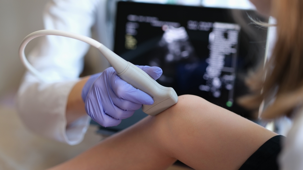
This is where emergency department ultrasound plays a vital role in patient care. These portable point-of-care devices have also become an indispensable tool at the bedside, empowering physicians to see beyond the surface.
Nevertheless, the journey towards integrating ultrasound imaging into the standard toolkit of the emergency department has been far from smooth. Despite the challenges, the applications for ultrasound in the emergency department continue to expand, bringing about a revolutionary and streamlined approach to delivering clinical care.
The journey to emergency department ultrasound
Although ultrasound was first used by shipbuilders to detect flaws in metal hulls, today the word conjures mental images of pregnancy scans.1 That's understandable - obstetricians were the first doctors to embrace this technology as a diagnostic tool because it does not expose a developing fetus to radiation.1
Before this, though, brain scans were the first medical application for ultrasound.2 These attempts were largely unsuccessful due to interference from the reflective skull surrounding the brain. Momentum picked up in World War II when British obstetrician Ian Donald used ultrasound to distinguish between benign and malignant masses.3 He and another physician, John MacVicar, later designed their own ultrasound machine that outperformed X-Rays and physical exams in correctly diagnosing ovarian cysts.3
Still, despite proof of efficacy, ultrasound struggled to gain its clinical footing. At the outset, the biggest obstacle was resistance from radiologists, who argued that non-specialists should not wield imaging equipment for fear of misdiagnoses.1 But there were also additional hurdles. Early machines were large and bulky, and patients were submerged in water so these machines could capture images. In these early days, ultrasound was useful and effective, but ultimately inconvenient.
In the early 1990s, however, the tides began to turn toward point-of-care ultrasound use. When treating trauma patients in the emergency department, surgeons at Grady Memorial Hospital recognized how quickly ultrasound provided accurate evaluations.1 Based on these observations, they introduced the Focused Assessment with Sonography in Trauma—the FAST exam, an assessment that is now a mainstay emergency department imaging test that quickly pinpoints any internal bleeding typically associated with a chest hemorrhage. FAST results frequently help emergency providers decide on treatment options or determine if additional imaging, such as a CT scan, is necessary.
The digitization of ultrasound
In addition to being large and difficult to maneuver, early ultrasound machines required patient transport, potentially wasting valuable clinical time. At the time, technologists needed specialty training to capture grainy images on Polaroid film.
Digitization changed everything, however. When silicon chips hit the market, ultrasound machine sizes shrank, and image quality improved.1 User interfaces were simplified, helping technicians to do their job more efficiently. And a mid-1990s grant from the Defense Advanced Research Projects Agency (DARPA), funded the creation of a portable ultrasound device that could be carried onto the battlefield—and into the emergency department today. In fact, digitization advances now mean physicians have access to ultrasound devices that can conveniently connect to smartphones or tablets.
Uses for ultrasound in the emergency department
Thanks to digitization, point-of-care ultrasound use is growing rapidly. In fact, many medical schools provide a handheld device for every first-year medical student.
Evidence already shows that using these devices can lead to faster, safer, more accurate, and more affordable patient care.4 Digital tools, such as the ViewPointTM 6 for Point-of-Care Ultrasound, can support encounter-based imaging workflows that can lead to more timely diagnosis and treatment. By using ultrasound to assess patients for organ damage, pneumonia, blood clots, detached retinas, eye bleeding, gout, and rheumatoid arthritis, physicians can help patients avoid unnecessary or more invasive procedures.
Alongside assessing patients with these problems, as well as trauma or traumatic injury, emergency department ultrasound can be useful in several other cases:
- Abdominal pain: For patients presenting with abdominal pain, point-of-care ultrasound can pinpoint kidney stones, abdominal aortic aneurysms, appendicitis, gallstones, or bowel obstructions.
- Shortness of breath: Point-of-care ultrasound can determine whether heart or lung problems are responsible for a patient's shortness of breath.
- Chest pain: Many patients come to the emergency department with a non-specific complaint of chest pain. Ultrasound can help quickly narrow down the cause.
- Shock: Emergency department ultrasound can identify the root cause and type of shock within six minutes.
- Cardiac arrest: Ultrasound can guide emergency providers through any resuscitation efforts.
- Intravenous (IV) lines: Ultrasound guidance can help providers accurately place IV lines in patients with difficult-to-find veins. Images also allow providers to specifically administer pain medication to targeted areas.
- Tuberculosis: Getting results through traditional tuberculosis testing can take several days. Emergency department ultrasound can detect tell-tale microabscesses and lymph nodes within minutes, accelerating treatment and recovery.
- Pre-operative/post-operative assessment: Using ultrasound before or after surgery can help clinicians and surgeons evaluate the patient's condition, guiding treatment and recovery decisions.
Ultrasound can also impact emergency care outside the hospital, particularly in rural areas where patients have limited access to medical services. For example, with patients who receive at-home care, clinicians can use the portable ultrasound to quickly assess any chest pain or shortness of breath. With this information in hand, providers can decide whether to send the patient to the emergency department or suggest medication or other treatment changes that can address the symptoms.
The Implications of Artificial Intelligence in Ultrasound
The integration of artificial intelligence (AI) into point-of-care ultrasound systems holds the potential to revolutionize accessibility and usability for emergency department healthcare providers without specialized imaging training. By leveraging AI algorithms, several significant implications emerge, paving the way for improved patient care and diagnostic accuracy.
Algorithms that can collect automatic measurements, such as how much blood the heart pumps per beat or the size of a lung nodule, may take on more administrative labor like making estimations or taking manual measurements to free up clinician time.6 By swiftly capturing this data, AI-powered ultrasound technology may expedite the detection and diagnosis of conditions like heart failure or potential lung cancer.
AI can also improve the performance of novice learner (NL) operators, or sonographers with minimal experience or suboptimal training. One study tested seven NL operators with limited to no prior POCUS experience for the ability to identify pneumonia in 32 pediatric patients. The study found that sensitivity, specificity, and accuracy of NL AI‐augmented interpretation were 66.7% (confidence interval [CI] 9.4–99.1%), 96.5% (CI 82.2–99.9%), and 93.7% (CI 79.1–99.2%).7 Another study tested an ultrasound lung imaging diagnosis model based on an artificial intelligence algorithm in neonatal respiratory distress syndrome (NRDS). The results showed that the segmentation accuracy of the algorithm was remarkably high, accurately extracting feature information from images.8
Furthermore, AI applications can offer valuable guidance during ultrasound scans, especially for novice users. Caption Health, recently acquired by GE HealthCare, has trained AI to recognize a diagnostic-quality cardiac image, and the software can provide directions on how to move or rotate the probe to acquire the optimal image. With clinical AI applications, ultrasound examinations can be easier and faster, lead to more precise diagnoses, and ultimately improve treatment decision-making.
Simple, Fast Solutions
As helpful as ultrasound is, in the ED, every second matters. Emergency care physicians need to make rapid decisions based on accurate and clear images. With auto tools, clinicians can save time and get the information necessary for physicians to take fast action. These grab-and-go tools can simplify complicated processes, such as shock or trauma assessment, by utilizing automated calculations that focus on indicators of the patient's condition, like the heart and lungs, and quickly gauging anything from internal bleeding to pneumothorax.
It's easy to think of ultrasound as a fundamental medical tool today, but its journey to clinical acceptance is now gaining substantial momentum. With continued digitization and integration of AI algorithms and tools that enhance encounter-based workflows, the value of adopting point of care ultrasound is becoming clearer, and the use of trauma ultrasound is becoming even more widespread. It's easy to see why, given the positive impact on patient and clinical outcomes.
REFERENCES:
1. Marks C. Could ultrasound replace the stethoscope? The New Yorker. January 20, 2023. https://www.newyorker.com/science/annals-of-medicine/could-ultrasound-replace-the-stethoscope. Accessed April 17, 2023.
2. The History of Ultrasound. BMUS. Accessed April 17, 2023. https://www.bmus.org/for-patients/history-of-ultrasound/.
3. Merz, Eberhard, David H Evans, Yi Dong, et al. History of Ultrasound in Obstetrics and Gynaecology from 1971 to 2021 on Occasion of the 50 Years Anniversary of EFSUMB. Medical Ultrasonography, 2022. https://doi.org/10.11152/mu-3845.
4. Hsieh A, Baker MB, Phalen JM, et al. Handheld point-of-care ultrasound: Safety considerations for creating guidelines. J Intensive Care Med. 37(9) (2022). doi:10.1177/08850666221076041.
5. ViewPoint 6 Point of Care Ultrasound. GE HealthCare. Accessed July 9, 2023. https://www.gehealthcare.com/products/ultrasound/ultrasound-reporting/viewpoint-6-pocus
6. Karami E, Shehata MS, Smith A. Semi-automatic algorithms for estimation and tracking of AP-diameter of the IVC in ultrasound images. J Imaging. 2019;5(1):12. (2019). doi:10.3390/jimaging5010012.
7. Nti, B., Lehmann, A. S., Haddad, A., et al. (2022). Artificial Intelligence-Augmented Pediatric Lung POCUS: A Pilot Study of Novice Learners. Journal of ultrasound in medicine : official journal of the American Institute of Ultrasound in Medicine, 41(12), 2965–2972. https://doi.org/10.1002/jum.15992
8. Wu, Y., Zhao, S., Yang, X., Yang, C., Shi, Z., Liu, Q., Wang, Y., Qin, M., & Zhang, L. (2022). Ultrasound Lung Image under Artificial Intelligence Algorithm in Diagnosis of Neonatal Respiratory Distress Syndrome. Computational and mathematical methods in medicine, 2022, 1817341. https://doi.org/10.1155/2022/1817341
ViewPoint is a trademark of GE HealthCare.
Product and features may not be available in all countries and regions. Full product technical specification is available upon request. Contact a GE HealthCare representative for more information.
JB25239XX