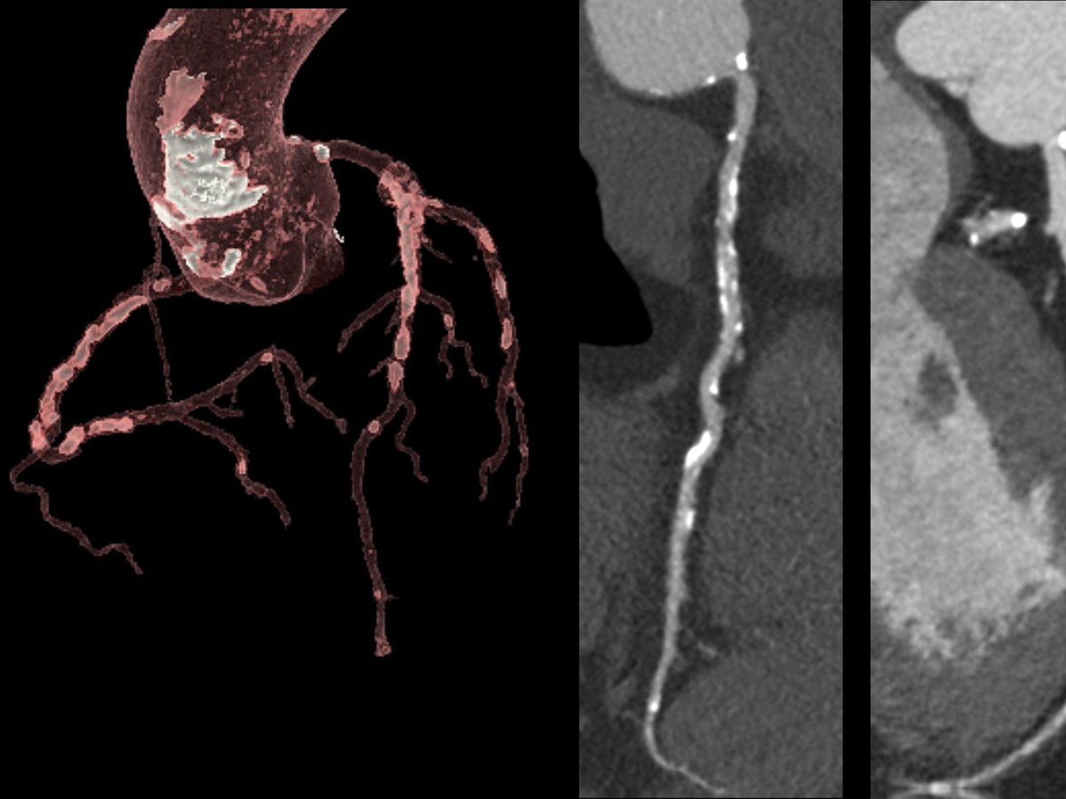
Over the past decade, advances in diagnostic imaging technologies have played a significant role in advancing personalized care and earlier disease diagnosis. These tools have specifically impacted diagnostic imaging in patients with cardiovascular disease.[1] Cardiovascular diseases remain the leading cause of death globally, with about 17.9 million lives taken each year, according to the World Health Organization.[2] Advanced imaging technologies, such as computed tomography (CT), have become invaluable for patient diagnostics, where clinicians rely on high-quality CT images to ensure diagnostic accuracy.
Cardiac CT imaging has transformed the detection, characterization, and stratification of coronary artery disease risk in individuals. A non-invasive imaging procedure, coronary computed tomography angiography helps identify plaque and blockages in the arteries and is used as a first-line test for patients presenting with chest pain. This procedure also informs clinicians for diagnosis and guides management strategy.[3] Every year, over 20 million U.S. patients present with chest pain, and estimates suggest that by 2025, 10–15 percent of all CT scans performed globally will be coronary CT angiography.[4]
Given the growing disease burden and subsequent need for imaging services, today’s healthcare professionals are facing higher expectations to serve patient communities with the latest in CT technology. Ongoing clinical pressures, such as complex imaging exams and a heavy patient load, require CT systems to meet a broad range of diagnostic needs. However, the financial and operational barriers to maintaining the most current technology are more prevalent than ever.
Optimizing asset management with scalable CT
Radiology administrators recognize the need to keep up with the latest medical imaging technology, boost productivity, and manage increasing demands for imaging. Accessing the latest technology and improving department workflow and productivity are high priorities for the next several years.[5]
Radiology departments are under pressure to maintain state-of-the-art imaging technology to serve their patient communities across every clinical application. When the pace of innovation is only getting faster, the challenge of technology obsolescence is also growing. Radiology departments worry that, in the not-so-distant future, a new system purchased today won’t be state-of-the-art for very long.
Industry partners like GE HealthCare are innovating in CT technology to generate high-quality images and create opportunities that make technology more accessible to more patients.
“We’re committed to developing and delivering the latest in CT technology to our customers, but also making it more accessible to democratize cardiology imaging,” said Samantha Barr, Senior Product Manager for Premium CT at GE HealthCare.
With a new CT platform developed by GE HealthCare, radiology departments can be “future ready” with built-in scalability and upgradability options.
“This powerful CT technology can adapt to the changing needs in cardiac imaging and across the radiology environment,” Barr continued. “We designed our new modular and scalable CT platform to handle complex protocols across anatomies, helping our customers achieve success today and also tomorrow.”
With its ability to scale detector coverage from 4, 8, or 16 cm of coverage through upgrades, the new CT platform delivers all the technology a customer may need today and can grow when needs change. The design has a common look and feel across its fleet and enables workflow optimization with effortless protocol standardization built in.
Improving operational efficiencies to streamline workflow and increase throughput
Intuitively designed systems and software solutions can help increase efficiency in radiology by using data and advanced technology to automate routine tasks, shorten acquisition times, streamline workflows, and prioritize urgent cases. However, implementing effective workflow solutions to accommodate all the necessary requirements for each exam has become more challenging with increasingly complex protocols.
Fortunately, the availability of artificial intelligence (AI) applications to aid in workflow optimization is quickly growing, leading to improvements in exam setup, protocoling, and patient positioning. AI-based tools are alleviating the time demands on radiology workflows, providing better standardization, and allowing radiology staff to spend more time on patient care. GE HealthCare’s CT platform uses AI and intuitive design to work in tandem with its workflow application, increasing efficiency in clinical workflow during the pre- and post-scan environments, as well as during the scan.
Bolstering clinical confidence in cardiac CT with advanced technology
CT technology is improving at a robust pace, leading to superior image resolution and fast volume coverage. Systems with an increased number of detectors and shorter gantry rotation times, as well as the advent of systems equipped with dual-source X-ray tubes, are enabling these advancements. Even with advances in CT technology, high-quality cardiac CT images often require more complex image acquisition protocols. The motion of the beating heart and patient health factors such as blood pressure and body weight also raise challenges.
The CT platform developed by GE HealthCare delivers powerful technology capable of high image quality in a low-dose environment, even for challenging anatomy such as the heart. Any heart and heart rate can be imaged within one beat and one rotation of its detectors with the fast rotation speed of 0.23 seconds per rotation and wide Z coverage of 160mm, coupled with SnapShot Freeze 2 motion reduction technology, potentially eliminating the need to administer beta-blockers prior to imaging.
“Today, with a 0.23-second rotation, we don’t administer beta blockers,” explained Dr. Joost Delanote, radiologist at AZ Sint-Jan in Brugge, Belgium. “We don’t monitor the heart rhythm in advance, even when patients have a very high heart rhythm. That’s a significant improvement between the past and today’s CT scans.”
The introduction and adoption of AI-based solutions are impacting radiology with image processing algorithms and tools to correct for motion artifacts. GE HealthCare’s CT image reconstruction algorithm applies deep-learning technology across the imaging dataset and in concert with the system’s fast rotation speed to obtain the highest quality cardiac images, while improving iodinated contrast volume, even in a low-dose environment.
Preparing for tomorrow with intelligent, future-forward CT today
Industry leaders such as GE HealthCare are aware of the challenges radiology departments face. They are delivering on their commitment to provide intelligent efficiencies and improve imaging access. GE HealthCare’s new developments in CT technology hardware and software allow clinicians agility and adaptability to meet the changing healthcare needs of today’s patients—and remain prepared as patient needs evolve in the future.
- Watch the on-demand presentation: ‘Changing the beat in cardiac care with Revolution™ Apex platform'
- Learn more about Revolution™ Apex platform
cardiac CT, computed tomography, scalable CT, advanced CT Technology
DISCLAIMER
Not all products or features are available in all geographies. Check with your local GE HealthCare representative for availability in your country.
REFERENCES
[1] Diagnostic and Interventional Cardiology. CT scans appear to dramatically improve diagnosis of heart disease. DIcardiology.com. Published 2015. https://www.dicardiology.com/article/ct-scans-appear-dramatically-improve-diagnosis-heart-disease. Accessed January 20, 2023.
[2] World Health Organization. Cardiovascular diseases. WHO.int. https://www.who.int/health-topics/cardiovascular-diseases#tab=tab_1. Accessed January 20, 2023.
[3] Kite TA , Ladwiniec A , Arnold JR , et al. Early invasive versus non-invasive assessment in patients with suspected non-ST-elevation acute coronary syndrome. Heart. 2022 Apr;108(7):500–506. doi: 10.1136/heartjnl-2020-318778.
[4] Channon KM, Newby DE, Nicol ED, et al. Cardiovascular computed tomography imaging for coronary artery disease risk: plaque, flow, and fat. Heart. 2022;108:1510-1515. doi: 10.1136/heartjnl-2021-320265.
[5] IMV Medical Information Division. The IMV 2019 global imaging market outlook report. IMVinfo.com. https://imvinfo.com/product/imv-2019-global-imaging-market-outlook-report/. Accessed January 20, 2023.


