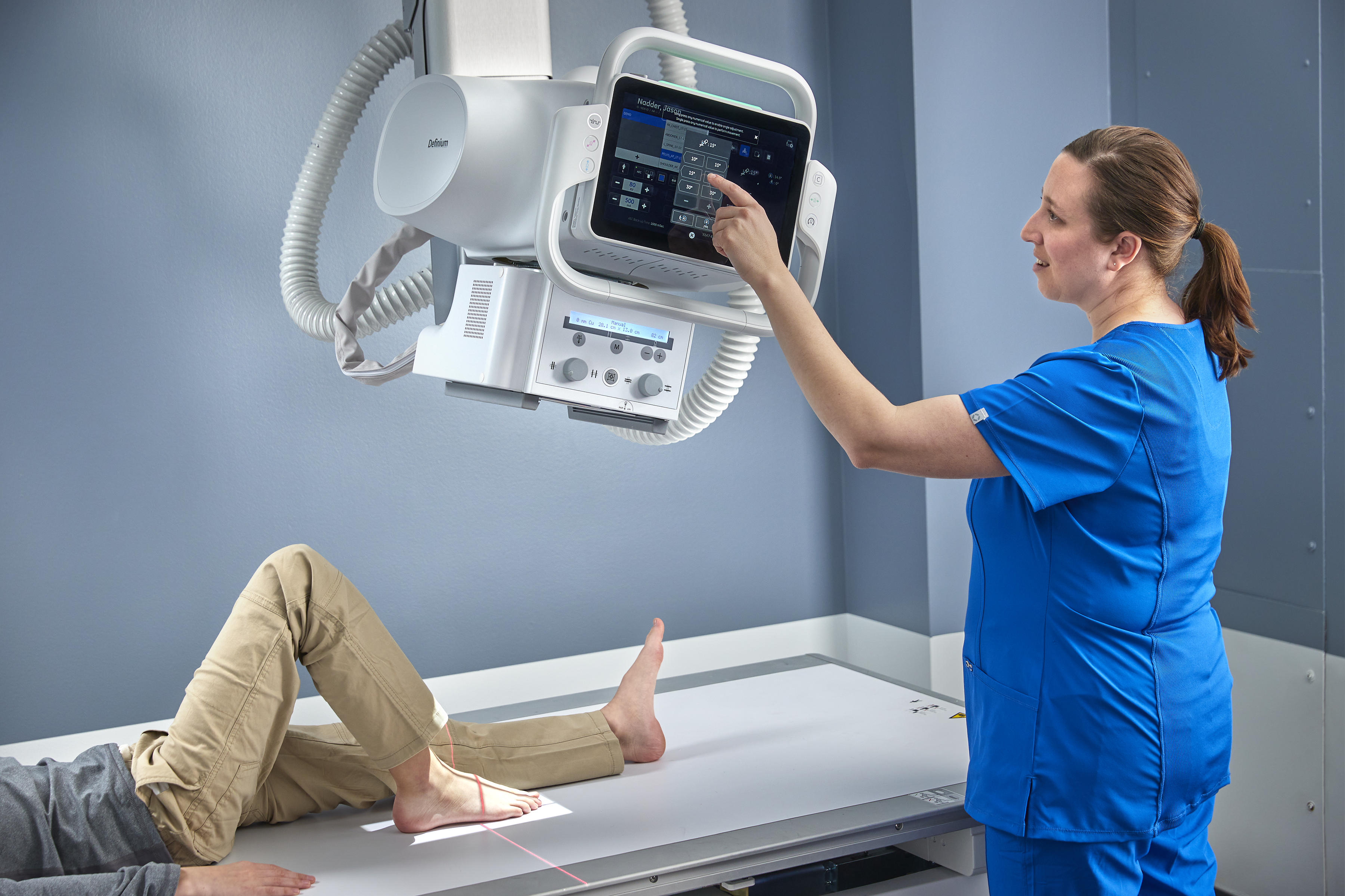
X-ray is the most utilized and most read imaging exam in radiology departments, accounting for 60 percent of all imaging studies.1 Because it is often the entry point to diagnostic imaging, getting the right image the first time is crucial.
Consider the potential benefits of obtaining a diagnosis from this first test without the additional cost, time, and resources created by additional imaging exams. Consider the potential benefits for both patients and staff.
Maintaining consistency in X-ray exam image quality across disparate patient populations
No two patients are alike; therefore, every exam is unique. Additionally, with different experience levels, imaging technologists likely perform the same exam differently. While beneficial for individualizing patient care, this variability can lead to inconsistent image quality, strain on technologists performing the exam, and can complicate reading for radiologists. In short, variability in the X-ray workflow can lead to operational inefficiencies that can reduce throughput and reduce capacity.
What if your X-ray system could help you do more by doing less? Streamlining your X-ray workflow leading to fewer repeat exams, fewer exam rejects, fewer patient positioning errors, and lower user variability that could lead to increased throughput and capacity.
GE Healthcare’s next-generation Definium™ 656 HD fixed X-ray, featuring the Intelligent Workflow Suite that includes Position Assist, Technique Assist and Patient Snapshot, assists technologists with consistent and efficient patient positioning and technique selection to help ensure that the first image counts each and every time. It’s a smart system that works to prevent errors before they occur.
Keeping an eye on the patient - improving the X-ray workflow
Up to 25 percent of exams can be rejected or repeated, with 68 percent of repeat exams due to poor positioning.2 Even with the most precise positioning, the patient may move. If the technologist is out of the room ready to capture the X-ray image, they may not notice misalignment until after the image is exposed, leading to repeat images.
With live streaming video and 3D depth cameras, the Definium 656 HD helps technologists keep their eyes on the patient even when they are not in the room. Mounted on the X-ray tube, a camera helps the technologist monitor the patient’s status, movements, and orientation before the X-ray is taken. It works anywhere the X-ray tube is pointed—for imaging at the wall stand, on the table, or with a wireless detector.
Position Assist goes one step further and overlays the detector boundaries, ion chamber locations and the active ion chambers right on the live video image of the patient using the smart camera. Position Assist updates in real-time to enable correct alignment regardless of the patient’s body size or position. With this real-time alignment information at the acquisition workstation, technologists can provide verbal instructions to the patient if they move, wait to capture the image until the patient moves back into position, or re-enter the room to reposition the patient. This information provides technologists with confidence that they can capture the image with correct positioning on the first take.
According to Jeannie Miller, Associate Radiology Director for the North Central Bronx Hospital, exam repeat rates have dropped from 4.5 percent to 1.2 percent thanks to the use of Intelligent Workflow Suite.
Personalized imaging with consistent quality
Using the 3D camera, Technique Assist measures the thickness of the patient and, using a database of programmable sizes, suggests a patient habitus optimized to the specific patient. It improves technique selection across 30 anatomy/view combinations including the abdomen, chest, pelvis and spine. Employing this feature results in more consistent imaging across the patient population and a reduction in variability across technologists, which also assists radiologists with confidence and efficiency when reading studies. For smaller patients, it can also potentially reduce their dose associated with overexposure.
Orlando Ortiz, MD, Chair of the Department of Radiology for the North Bronx Healthcare Network and a department radiologist, says, “In terms of the image consistency, it has been very good. The area of coverage, the collimation, the technique, the appropriate penetration for the specific studies—whether they are chest, abdomen, extremities, etc.—have been really spot on and steady. It’s high quality on a consistent basis.”
Further improving clinical confidence for radiologists is Patient Snapshot. Patient Snapshot takes an optical picture simultaneously with the X-ray exposure and can attach it to the study, so radiologists have more context regarding external objects, positioning limitations and patient orientation, and any deviations from ideal imaging conditions. With heavy reading workloads and time pressures contributing to burnout among radiologists,3 any information that can reduce questions from radiologists and prevent slowing down diagnoses can improve efficiency and clinical confidence.
With the Definium 656 HD and the Intelligent Workflow Suite, it’s almost like every technologist has a personal assistant to help them get the right image the first time. And there is more to come. This is only the beginning of GE’s vision to deliver the tools and technologies that can assist technologists and radiologists in their daily workload routine.
REFERENCES
- IMV 2019 X-ray CR / DR Market Outlook Report) page 9, 37
- Little, Kevin J., et al. "Unified database for rejected image analysis across multiple vendors in radiography." Journal of the American College of Radiology 14.2 (2017): 208-216.
- Chetlen AL, Chan TL, Ballard DH, et al. Addressing Burnout in Radiologists. Acad Radiol. 2019 Apr;26(4):526-533.

