When Jill Sievers went for her annual mammogram in March 2018, a mammographer handed her a pamphlet about automated breast ultrasound (ABUS), a screening test for women with dense breast tissue.
A follow-up ultrasound that doctors ordered because of her inconclusive mammogram also found nothing malignant, just a few cysts. Days later, Sievers happened to have her annual exam scheduled with her OB-GYN, so she mentioned the ABUS pamphlet and requested that she be scanned with the new technology, because she knew that she had dense breasts. Sievers’ doctor agreed that it was appropriate and sent her for the exam that afternoon.
Within two days, Sievers was diagnosed with breast cancer.
“I’ve always gone for yearly breast cancer screenings and as usual had regular and 3D mammography,” says Sievers, 61, of Hiawatha, Iowa. “My cancer wasn’t found with either of these imaging tools or a follow-up targeted breast ultrasound. Only the ABUS image showed my tumor.”
GE Healthcare’s ABUS is the first FDA-approved screening ultrasound technology that is designed to pinpoint cancer in dense breast tissue. It’s intended to be used in conjunction with mammography because each tool assesses breast tissue differently.[1] 3D mammograms are considered tomography, or three-dimensional views of a breast based on a series of low-dose X-ray images, while ABUS technology uses ultrasound imaging to create views of the interior of the breast. Women who receive ABUS, which the FDA approved in 2012, lie flat on a table while a scan head is placed over the breast. The 15-centimeter-wide, field-of-view transducer scans the entire breast tissue, which facilitates more confident readings in multiple planes and volumes. Compression levels can be selected, which makes the exam as comfortable for women as possible.
“It’s not a substitute for tomography, because they’re complementary, but there are additional ways of looking at breast tissue other than using X-rays ... and that parlays into a potential earlier diagnosis,” says Arnold Honick, M.D., a Cedar Rapids, Iowa, diagnostic radiologist who identified Sievers’ breast cancer. “Even if you go back and look at your mammogram, you can’t see the cancer that was detected by ultrasound.”
Dr. Arnold Honick
Sievers’ early diagnosis caught her breast cancer before it spread to the lymph nodes. It also enabled her to have a specific type of reconstructive surgery that would not have been possible if the tumor had been larger. She was able to avoid radiation therapy, which may not have been an option if she had been diagnosed during her annual mammogram the following year.
“Who knows how much it would have grown or become more conspicuous?” Honick says. “[It] likely would have been at a larger, advanced stage and affected her prognosis.”
Help for women with dense breasts
About 40% of women of mammography age have dense breast tissue — in Asia the figure is around 70%[2] — which can both hide (or mask) cancer on a mammogram and increase the risk of developing breast cancer. Breasts contain a mix of glandular tissue, fibrous connective tissue and fatty tissue. Dense breasts are composed of more glandular and fibrous connective tissue than fatty tissue. Only a mammogram can detect breast density, and while breast density reporting/education varies across the United States, currently 38 states and the District of Columbia require some level of breast density notification after a mammogram.
“If you took the patients with the very highest breast density — those ones that are extremely dense — and compare them to patients that have fatty breast tissue, they have a four to six times greater risk of breast cancer,”[3] Honick says.
Additionally, dense breast tissue may obscure up to half of cancers on mammograms among women with extremely dense breasts.[4] ABUS screening alone improves invasive cancer detection over mammography by 35.7%,[5] finding hidden cancers among women with dense breasts.
ABUS, an automated procedure, provides consistent results that may be enhanced by AI Assistant, which integrates intelligent algorithms powered by the FDA-approved third-party tools QVCAD and KOIOS DS Breast to assist in detecting and characterizing breast lesions.
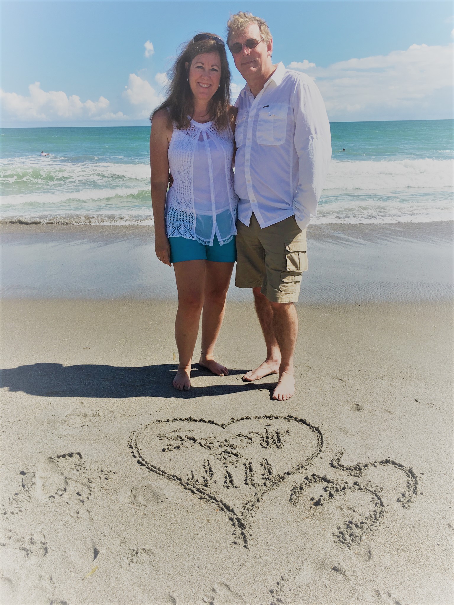
Jill Sievers with husband Scott, celebrating their 35th wedding anniversary.
“It can speed up your ability to interpret the exam and also potentially assure that you’re not going to overlook something,” Honick says. “The biggest benefit to me has been the efficiency of reading [or] interpretation.”
ABUS screening may be crucial for younger women, who are more likely to have dense breasts.[6]
“The younger you are, the more common that breast cancer is going to be a more aggressive type, so therefore it’s even more important to try to detect it at an early stage,” Honick says.
A powerful exam for women to request
Women as young as age 40 may be advised to schedule annual mammograms.[7] It’s also essential for women to learn whether they have dense breasts so they can seek ABUS screening as needed to search for hidden cancers that mammography alone cannot reveal. While reimbursement coverage differs from jurisdiction to jurisdiction, ABUS screening is widely available in most geographies around the world.
Four years after her cancer diagnosis and treatment, Sievers couldn’t be happier that she advocated for her health by requesting ABUS. Since her ABUS cancer diagnosis and double mastectomy, she has been able to see her son get married and three of her five grandchildren receive their First Communion. She has also been able to maintain her active lifestyle — doing aerobics, lifting weights, traveling, skiing, swimming and gardening. Sievers is grateful to her radiology team and thrilled to be living life cancer-free.
“They gave me that ABUS pamphlet that informed me of this amazing technology that was my golden ticket — I knew I had dense breast tissue, and I knew that reading dense tissue is a challenge,” she says. “Why put yourself through worse outcomes and a path with more procedures? Requesting ABUS makes the whole journey easier by finding cancer earlier, when less treatment is likely to be needed, and the imaging is totally pain-free and comfortable — a great golden ticket!”
REFERENCES
[1] GE Healthcare, Invenia ABUS, https://www.gehealthcare.com/products/ultrasound/abus-breast-imaging/invenia-abus.
[2] Etta D. Pisano, M.D., et al. “Diagnostic Performance of Digital Versus Film Mammography for Breast-Cancer Screening,” New England Journal of Medicine 353 (October 27, 2005): 1773-83.
[3] Norman F. Boyd, M.D., et al., “Mammographic Density and the Risk and Detection of Breast Cancer,” New England Journal of Medicine 356 (January 18, 2007): 227-36.
[4] Thomas M. Kolb et al., Comparison of the Performance of Screening Mammography, Physical Examination, and Breast Us and Evaluation of Factors That Influence Them: An Analysis of 27,825 Patient Evaluations,” Radiology 225, no. 1 (October 2002): 165-75.
[5] FDA, “Summary of Safety and Effectiveness,” PMA P110006 (2012).
[6] CDC, “What Does It Mean to Have Dense Breasts?,” last updated September 2021, https://www.cdc.gov/cancer/breast/basic_info/dense-breasts.htm.
[7] American Cancer Society, “American Cancer Society Recommendations for the Early Detection of Breast Cancer,” last updated January 2022, https://www.cancer.org/cancer/breast-cancer/screening-tests-and-early-detection/american-cancer-society-recommendations-for-the-early-detection-of-breast-cancer.html


