Precision and speed are part of the job description for anesthesiologists. Whether they're performing a nerve block or preparing for emergency surgery, they need a crisp, detailed image of what's happening inside their patients—ideally in real time. That's why ultrasound for anesthesia is quickly becoming a common standard of care in hospitals around the world.
Until recently, ultrasound was mostly used by radiologists and cardiologists, but the emergence of Point of Care Ultrasound (POCUS) has made this imaging technology more cost-effective and easier to use, and thus more accessible to anesthesiologists and other subspecialists. Over the past decade, anesthesiologists have identified many potential use cases for ultrasound in anesthesiology, and research has confirmed its many benefits for both real-time guidance and clinical decision-making.
Ultrasound for Anesthesia: Use Cases
Leading anesthesiologists and certified registered nurse anesthetists (CRNAs) have been using ultrasound for years, but as POCUS becomes more affordable and portable—and thus more pervasive within the specialty—governing boards and organizations have begun to weigh in on potential use cases and best practices.1 Now, residency programs are required to teach ultrasound in anesthesiology, and the American Society of Anesthesiologists (ASA) introduced POCUS guidelines and a certification program. This provides clinicians with qualifications in three primary areas: cardiac, lung, and abdominal ultrasound as well as six secondary applications: musculoskeletal/soft tissue, airway, ocular, transcranial Doppler, renal/genitourinary, and deep venous thrombosis.
Ultrasound-Guided Regional Anesthesia
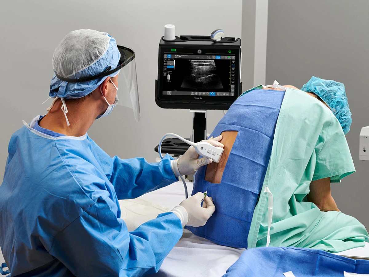
Ultrasound-guided nerve block is among the most common uses of POCUS in anesthesiology. To control pain during operations and other invasive procedures, anesthesiologists and CRNAs inject analgesic drugs as close to local nerve regions as possible, ideally without touching nerves or puncturing veins. Ultrasound-guided needle placement lets them visualize nerve regions in real time, so they can identify where to place the needle and continuously view where the needle is located in relation to nearby nerves and vascular structures, reducing the risk of possible nerve damage or accidental venous puncture.
Vascular Access
Ultrasound has also been shown to offer excellent guidance for difficult venous access. Anesthesiologists traditionally rely on physical landmarks to obtain vascular access, but this technique can be challenging with patients who are obese, have certain chronic medical conditions, or have vein degeneration due to age, illness, or intravenous drug use. Ultrasound lets anesthesiologists identify and access veins more easily and reduces the risk of accidentally puncturing unintended veins or arteries. It can also be used to screen for and identify deep venous thrombosis (DVT) that could form into a pulmonary embolism.
Airway Ultrasound
When an endotracheal tube is accidentally placed into a patient's esophagus or bronchi, it can cause neurological damage and death. Anesthesiologists can typically see where to place the tube without imaging, but in patients with a difficult airway, neck ultrasound provides an accurate visualization of the trachea and esophagus and reduces the risk of accidental esophageal intubation.
Surgical Risk Assessment
Ultrasound can help anesthesiologists identify cardiac, pulmonary, and gastric risk factors before and during surgery. For example, cardiac ultrasound can show valvular abnormalities, biventricular function, pericardial tamponade, blood volume, and cardiac ischemia. Pulmonary ultrasound can be used to assess patients for potential hypoxia, pulmonary edema, pneumothoraces, and lung consolidation. Gastric ultrasound is a quick and reliable way to scan a non-fasting patient's stomach contents to determine their risk for aspiration under general anesthesia during emergency surgery.
Benefits of Ultrasound for Anesthesia
There are many diverse use cases for POCUS in anesthesiology, and a growing body of research shows that it boosts efficiency, improves patient outcomes, and enriches quality of care.
Compared to other initial bedside assessment tools, ultrasound often costs less and while offering valuable diagnostic insights. For example, airway POCUS has experienced dramatic innovation over the past few years in identifying misplaced intubations. POCUS is also a fast and accurate way to detect pneumothorax, pleural effusions, and consolidation in critical patients instead of relying solely on chest auscultation or chest radiograph.
Proficiency with POCUS also enables ultrasound-guided regional anesthesia, which can improve patient outcomes with perioperative pain management and postoperative recovery. Compared to patients who receive general anesthesia (sedation) during surgery, patients who undergo ultrasound-guided regional anesthesia tend to recover more quickly with fewer complications.3 Compared to other nerve identification methods used for regional anesthesia, ultrasound guidance has been shown to increase the probability of successful nerve block, decrease needle placement time, and improve the overall safety of the procedure.
Challenges of Ultrasound for Anesthesia
POCUS is becoming an increasingly useful tool for anesthesiologists and CRNAs, but one limitation of the technology is that image quality and diagnostic accuracy depend on the skill and experience of the practitioner.2 Ultrasound-guided regional anesthesia includes the acquisition of acceptable ultrasound images of the nerve while avoiding artifacts, and needle artifacts may cause confusion and error during ultrasound-guided nerve blocks. However, acoustic artifacts are usually the result of incorrect assumptions during processing by the instrumentation. Physiological and pathological factors attributable to the patient can also affect image quality and interpretation.
Historically, anesthesiologists have not been extensively trained to use POCUS, but that has begun to change over the past few years. For example, the Accreditation Council for Graduate Medical Education (ACGME) recently updated its program requirements for anesthesiology residency programs to include ultrasound training, and the American Board of Anesthesiologists (ABA) now includes POCUS on board exams.1 Similarly, the European Society of Anesthesiology and Intensive Care has created POCUS guidelines for the perioperative use of regional anesthesia and vascular access, and the International Anesthesia Research Society now offers self-study POCUS webinars.4,5
At the same time that anesthesiologists and CRNAs are becoming increasingly proficient with POCUS, the technology is becoming more advanced—paving the way for even more diverse utilities and possibilities for ultrasound in the future of anesthesiology.
References:
1. Resources from ASA committees. American Society of Anesthesiologists. https://www.asahq.org/standards-and-guidelines/resources-from-asa-committees#POM. Accessed April 24, 2022.
2. Heinz, ER and Vincent A. Point-of-care ultrasound for the trauma anesthesiologist. Current Anesthesiology Reports. Jan 2022;1-9. https://www.ncbi.nlm.nih.gov/pmc/articles/PMC8771171/.
3. Zhao Y, Huiwen Z and Song M. Clinical observation of ultrasound-guided nerve block anesthesia on postoperative pain control of fracture patients. Journal of Healthcare Engineering. April 2022. https://www.ncbi.nlm.nih.gov/pmc/articles/PMC9010165/.
4. Boselli E, Hopkins P, Lamperti M, et al. European Society of Anaesthesiology Guidelines on peri-operative use of ultrasound for regional anaesthesia (PERSEUS regional anesthesia) Peripheral nerves blocks and neuraxial anaesthesia. Eur J Anaesthesiol. 2020;37:1–32. https://www.esaic.org/uploads/2021/01/perseus-part-ii-nerve-blocks-and-neuraxial-anaesthesia-november-2020.pdf.
5. International Anesthesia Research Society. IARS virtual education: research series. https://iars.org/iars-webinar-series/.