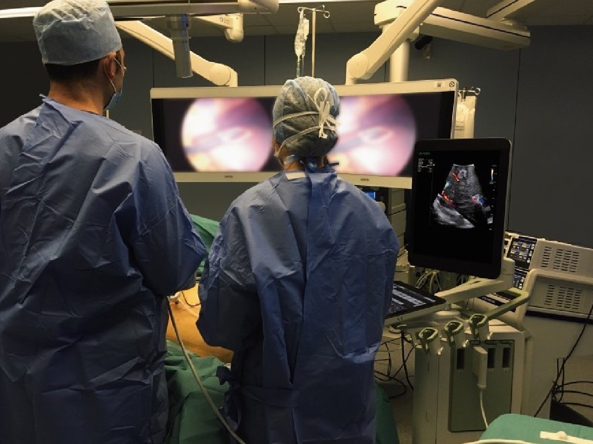
Taking cancerous tumors out of kidneys without damaging these vital organs was once a fantasy. Removing large portions or even the entire kidney using open surgery was the norm. Then, in 1991, came the first successful attempt to remove a kidney tumor using minimally invasive techniques. Today, what’s known as robotic partial nephrectomy — using cameras and robotic tools to remove precise portions of the kidney through tiny incisions in the abdomen — is the standard of care. Because surgeons can leave as much of the kidney intact as disease or injury will allow, patients can go on living with more kidney function, meaning less chance of kidney failure or need for lifelong dialysis treatments.
Now the new frontier in complicated surgeries like partial nephrectomy is intraoperative imaging: using surgical ultrasound and other technologies to peer inside the patient’s body during surgery to help guide doctors as they make incisions, position instruments, and confirm results. Intraoperative imaging has been shown to change how surgeons operate and alter the decisions they make mid-surgery. One such technology that’s favored by clinicians in the OR, including those who perform partial nephrectomies, is the bkActiv ultrasound system from BK Medical, a Boston-based ultrasound innovator that GE HealthCare acquired in 2021.
This week, GE HealthCare announced that bkActiv will expand to many more types of procedures in the operating room than before, including urology, colorectal, and pelvic floor procedures. The technology gives clinicians visual information that guides them during procedures and aids them in making informed, critical decisions. These insights can be especially useful in the growing field of minimally invasive and robotic surgery, designed to cause less pain and trauma, reduce complications, and send patients home faster.
“By expanding the bkActiv system, we are giving physicians the tools they need to provide precise treatment with the ability to see fine details in image guidance, which ultimately may allow for better outcomes for people who require urology, colorectal, and pelvic floor procedures,” says Urvi Vyas, general manager of surgical visualization and guidance–ultrasound at GE HealthCare.
Many ultrasound systems serve a general purpose. A clinician applies a probe, the instrument that generates the sound waves whose echoes produce the image, to a patient’s skin to get an (often fuzzy-looking) image, perhaps of a developing fetus. A trained radiologist who specializes in reading ultrasound typically interprets the images. By contrast, bkActiv was designed with surgeons and surgery in mind and includes a range of features that make it better suited to the unique environment of the operating room, says Adam Melnick, global product strategy director at GE HealthCare.
The bkActiv system is smaller and more mobile, easier to clean (with a touch-screen user interface), and much simpler to use, so that a surgeon can operate it with one hand while performing surgery with the other. Using specially designed probes designed to reach and image specific anatomy such as the prostate and the brain from inside the body, bkActiv produces images that are nothing like those fuzzy pictures typical of conventional ultrasound. Images are further enhanced using computer algorithms and computational models developed over decades, says Melnick.
Ultrasound in the OR is often used to identify and characterize cancerous lesions, especially in the kidneys, liver, and pancreas, but is showing up in ever more scenarios as the technology gains traction. The tricky practice of hunting cancer within the prostate is a good example, says Melnick. Most cancers are localized to a specific U-shaped band at the posterior of the prostate, a dense gland shaped somewhat like an inverted cone. The bkActiv system can highlight specific parts of the prostate for the surgeon and even illustrate the blood flow within them, something few other ultrasound technologies can match, making it easier to figure out where cancer is hiding.
“What image quality gets you is it just makes you more confident in what you’re seeing,” says Melnick, “and that helps guide you in your intervention.”
The expansion of bkActiv to more OR procedures will at first benefit the technology’s most enthusiastic adopters. Urologists, who specialize in treating diseases of the kidneys, prostate, and urinary tract, have been quick to adopt robotic surgery techniques as well as intraoperative imaging to assist with procedures such as prostate biopsies. With this change, more of their work can take place in the OR using the bkActiv system, meaning they can use their preferred tool to do more in one location, allowing them to better serve patients.
An emerging set of procedures called focal therapies provide an example of how bkActiv may be able to play a more prominent role. Removing the entire prostate because of cancer is fairly common today, says Melnick, but it comes with a host of complications, including erectile dysfunction and incontinence, that affect a patient’s quality of life after the surgery. Newer techniques involve directing energy at cancer cells to ablate, or destroy, a tumor. By placing bkActiv’s specially designed probes in specific positions around the prostate, surgeons can better aim the ablative energy so that it targets cancerous cells but leaves healthy ones intact.
The field of intraoperative imaging is evolving, and real-time, surgical-guided ultrasound is one of the fastest-growing technologies to enhance other imaging modalities used in the OR. MRI and CT taken pre-operatively are still preferred by some specialists, such as neurosurgeons, says Melnick. But he thinks surgical ultrasound has a strong case to complement them. Ultrasound provides active imaging and is easier for non-radiologists to use during surgery, and the equipment is smaller. Ultrasound utilizes sound waves instead of ionizing radiation, making it suitable for any patient, without the need for patient preparation before imaging.
GE HealthCare has been pioneering the use of ultrasound in medicine since the first systems appeared in the 1950s and ’60s, building on engineering breakthroughs such as the hydrophone and sonar, which allowed sailors to hunt for submarines early in the 20th century. GE HealthCare acquired BK Medical in order to complement its existing portfolio of ultrasound technologies, providing more options to clinicians in more settings.
“Adding the fast-growing and relatively new field of real-time surgical visualization to GE HealthCare’s pre- and post-operative ultrasound capabilities will create an end-to-end offering through the full continuum of care,” said Roland Rott, president and CEO of GE HealthCare Ultrasound, when the deal for BK Medical was announced in 2021. “GE HealthCare and BK Medical share a passion for clinical innovation, and I’m excited to welcome BK Medical to our team.”


