Digital technology is present in nearly every aspect of everyday life and has fundamentally altered the way people live. The same can be said about the effects of shifting to digital technology in medical imaging—it has altered the practice of medicine by exponentially improving the power of imaging diagnostics. Some of the biggest transformations are being made in molecular imaging (MI) technologies. Early on, leaders in MI brought clinicians hybrid imaging technology that combined the localization capabilities of computed tomography (CT) with the functional information from single photon emission tomography (SPECT) and subsequently from positron emission tomography (PET). With the introduction of solid-state detector technologies, innovators continued to push the limits of medical imaging and enabled clinicians to visualize disease with finer detail and with higher resolution. These new imaging systems introduced more imaging data for processing as well as highly sophisticated automated tools and AI-based reconstruction algorithms to assist clinicians as they rendered complex diagnoses.
Innovation continues to propel MI forward with the newest breakthrough technology in SPECT/CT, StarGuide™. Developed by GE Healthcare, StarGuide’s innovative design, cadmium zinc telluride (CZT) detectors, and intuitive workflow to automate patient positioning are making quantitative SPECT/CT achievable in routine clinical practice and advancing efforts enable precision healthcare and more personalized medicine for patients.
Making the case for quantitative SPECT/CT
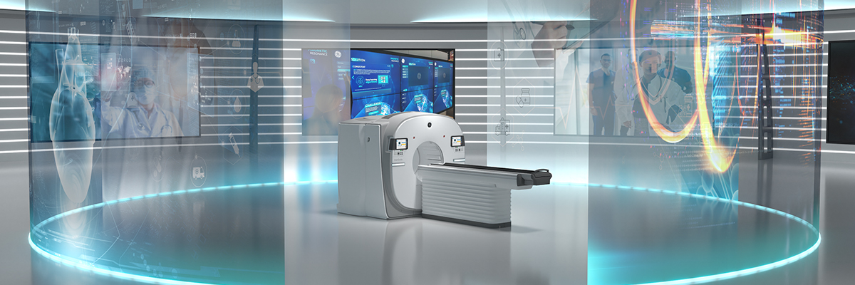
The digital SPECT/CT system is based on a completely new design with a shape-adaptive gantry and intuitive workflow. Once the patient is on the table, Optical Scout quickly maps the patient so the detectors and table automatically position themselves for close proximity and contactless scanning. Truly a next generation SPECT/CT, there are 12 CZT Digital Focus Detectors that surround the patient scanning them in 3D, minimizing distance from the patient across a variety of body shapes when scanning areas such as the spine, heart or brain. The system is also uniquely capable of scanning in dual-isotope mode, so clinicians can visualize multiple tracers simultaneously due to the excellent energy resolution of CZT.
Barry Siegel, MD, Professor of Radiology and Chief of the Division of Nuclear Medicine at the Washington University School of Medicine in St. Louis, Missouri, and the first clinical user of General Purpose (GP) SPECT/CT CZT detector technology in the US said that CZT’s capability to improve spatial and energy resolution also contributes to quantitative accuracy and should be considered vital to the adoption of quantitative SPECT/CT. He also believes that the incorporation of sophisticated AI-based reconstruction software is equally important.
“This detector technology can meet the challenges associated with designing the next generation of acquisition hardware,” Dr. Siegel explained, “however, the need for innovation cannot stop there. We need to incorporate new AI-enhanced applications to assist with a multitude of tasks such as relief from various laborious practices of lesion segmentation, longitudinal comparisons and absorbed dose calculations. We must have this technology applied to our MI space to compliment the CZT acquisition so reading SPECT/CT compared to planar images can be comparably efficient, making the case for quantitative SPECT.”
New CZT-based MI technology can propel the possibility of leveraging digital SPECT to enable next level personalized, precision healthcare through quantitative imaging, accurate dosimetry, and the application of new theranostic pairs of imaging and therapeutic radiopharmaceuticals.
“The possibility of whole-body dynamic SPECT/CT is exciting, indeed, but to make quantitative SPECT commonplace, the nuclear medicine community will need to collect the evidence to show that these efficiencies are possible and prove they lead to improved patient outcomes,” added Dr. Siegel.
Evaluating effective clinical applications of digital SPECT/CT
The new digital SPECT/CT system, StarGuide, was first installed at Centre Hospitalier Régional d'Orléans, in France, where the nuclear medicine team uses it in daily clinical practice for studies in bone imaging, cardiology, oncology and neurology. They feel that it will further propel the SPECT modality to help them deliver on the promise of nuclear medicine and precision health. Likely to become a major part of nuclear medicine in the near future, digital CZT detector technology capitalizes on improved sensitivity, spatial and energy resolution coming together, and not at the cost of one for the other as seen in systems built with sodium iodide (Nal) scintillation detectors.
They’ve also used the system to image multiple isotopes simultaneously. To date, the Hospital of Orléans team has scanned more than 300 patients and is delighted with the system’s efficient workflow and imaging capabilities and is optimistic about its potential expansion to new clinical areas and to support theranostic efforts in patient care.
Installed as a GP camera in the nuclear medicine department, StarGuide has been utilized primarily for bone studies, according to Matthieu Bailly, MD, Head of Nuclear Medicine at the Orléans facility, but has also been evaluated in other clinical applications.
“Our procedure mix is about 70 percent bone exams,” explained Dr. Bailly. “Bone scintigraphy is a major part of any nuclear medicine department, but we’ve also evaluated the system in other areas, such as whole-body studies, brain perfusion and myocardial perfusion with impressive results.”
The combination of digital SPECT imaging with CT also enabled the team to explore certain clinical applications with more detail.
“This technology represents a breakthrough in the world of SPECT imaging,” explained Dr. Bailly. “Using its innovative design and 3D ring CZT detection, we are now able to acquire full SPECT images, and the improvements gained in image quality and resolution will help us to improve our nuclear medicine procedures. The new system provides impressive results in cardiac imaging, for example. The focus mode allows us to accurately acquire any type of patient, including obese patients. And we’re thinking about adding the CT for attenuation correction, and calcium scoring to our myocardial perfusion imaging. These can help to improve the overall diagnostics. In bone SPECT, we hope to use the focus mode to explore bone assessment because we’re able to generate information and anatomical details for spine and extremities that isn’t available using conventional SPECT/CT.”
Gaining operational efficiencies through digital SPECT/CT automation and design
The system’s 12 CZT focus detectors automatically move to acquire data in close proximity to the patient. Infrared sensors guide the detectors and accurately center the patient in the gantry, a task that, on systems without this automation, could require several attempts and the technologist moving from gantry to workstation to see that the patient is positioned properly and could result in error. This new workflow efficiency has not gone unnoticed by the Hospital of Orléans team.
“It really simplifies patient positioning,” said Pierrick Oudot, Nuclear Medicine Technologist at the Orléans facility. “Centering the patient and starting an exam is very efficient. The system proceeds automatically based on the patient’s body contour. The detectors are always as close as possible to the patient which improves spatial resolution and sensitivity. The optimized mechanical movements of the detectors contribute to shorter acquisition times, making the exam more tolerable by the patients.”
The system’s unique ability to simultaneously image multiple tracers in a single scan helps to reduce the need for multiple patient visits, and in some cases, may help avoid additional patient sedation.
Advancing the future of molecular imaging with 3D dynamic SPECT/CT
The innovations in digital SPECT/CT imaging support many new paths in quantitative imaging and theranostics, as well as precision health and individualized patient care. As more clinical applications are evaluated and reported, 3D dynamic SPECT/CT[1]may contribute to the increasing diagnostic power of molecular imaging and be routinely used to provide clinicians with the data they need to help them make personalized care decisions and treatment recommendations for optimal patient outcomes.
For more information about GE Healthcare’s StarGuide™ SPECT/CT, click here.
To visit GE Healthcare virtual experience at the SNMMI Annual Meeting, register and login here.
View the on-demand replay of Nuclear medicine scanners: A journey from the past to the future to learn more about StarGuide.
[1] “Acquisition Only: The acquired dynamic data is stored in a list file.”


